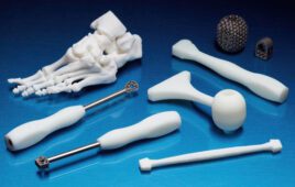
[Image from University of Minnesota]
1. 3D printed models help separate conjoined twins
About three months into her pregnancy, Paris Bryan found out that her twins were thoracoomphalopagus conjoined – from the chest to the belly button. The twins didn’t have a breastbone and their livers were fused and their hearts were separate, but touching.
Surgeons, cardiologists and other medical experts prepared for the twins arrival to carefully separate the twins.
“We developed a clear-cut plan for them. We started at the beginning with prenatal care, to delivery, to post-delivery care and finally the separation surgery and then post-separation care,” said pediatric surgeon Daniel Saltzman, chief of pediatric surgery at the University of Minnesota Masonic Children’s Hospital, in a press release.
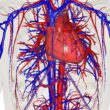
Saltzman and his team created a 3D model of the babies’ hearts before the separation surgery with help from the Medical Devices Center at the University of Minnesota. The model was able to show doctors a connection between the hearts. Because of this discovery, pediatric cardiothoracic surgeon Tony Azakie suggested a different surgical approach that could separate the hearts as well.
They separated the chest and fused livers first then separated their connected hearts. The hearts started beating separately.