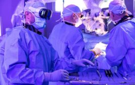
Image courtesy of MARS Bioimaging
New Zealand scientists performed the first-ever 3D, color X-rays on a human, using technology that could improve medical diagnostics in oncology, cardiology, neurology and orthopedics.
Based on traditional black-and-white X-ray technology, the scanner incorporates the Medipix3RX detector chip, a particle-tracking technology developed for the CERN Large Hadron Collider. It was developed by the Medipix3 Collaboration, which comprises CERN in Geneva and 18 other research institutions worldwide.
The scanner records the energy of each photon as it collides with pixels while the shutter is open, allowing for high-resolution, high-contrast pictures. It then uses computer algorithms to reveal the density of different materials, such as the percentages of water, calcium and fat in human tissue, explained Anthony Butler, a medical radiologist who worked on developing the scanner with his father, Phil Butler, a physics professor at the University of Canterbury in Christchurch, N.Z.
The machine’s “small pixels and accurate energy resolution meant that this new imaging tool is able to get images that no other imaging tool can achieve,” Phil Butler said in a prepared statement.
In preclinical use on animals at universities in New Zealand, Europe and the United States, researchers have been able to identify different cell lines in cancerous tissue, Anthony Butler said in a phone interview. The scanner could also be used to non-invasively measure arterial plaque to help diagnose the risk of heart attack or stroke as well as to track patients’ progress following treatment, he added. Researchers have also been using the scanner to study bone diseases such as arthritis and osteoporosis.
A clinical trial of orthopedic and rheumatology patients in New Zealand is planned for the coming months. According to the CERN, the images very clearly show the difference between bone, muscle and cartilage, but also the position and size of cancerous tumors, for example.
“This color X-ray imaging technique could produce clearer and more accurate pictures and help doctors give their patients more accurate diagnoses,” said a CERN statement.
The technology is being commercialized by New Zealand company MARS Bioimaging, launched by the universities of and Canterbury and Otago, which helped develop the scanner. MARS has a prototype human system running in New Zealand. The scanner has been in development for about a decade.
“It’s no good just doing science,” Anthony Butler said. “You’ve got to produce a product for people to use.”