PINE BROOK, N.J., Aug. 2, 2011 /PRNewswire/ — A new visual
evoked potential method has been developed to objectively assess
visual field defects in patients with ocular and/or neurological
conditions. The research team of Kenneth Ciuffreda, OD, PhD, Diana
Ludlam and Naveen Yadav at the SUNY State College of Optometry,
Department of Vision Sciences (New York, NY) presented a scientific
poster called “Effect of Different Stimulus Configurations on the
Visually Evoked Potential (VEP)” at the 2011 Association for
Research in Vision and Ophthalmology (ARVO) Annual Meeting. The
purpose of the study was to assess the effect of different stimuli
on VEP results, with the future goal of using the method as an
objective form of visual field testing.
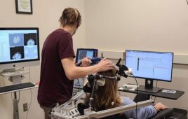
Traditionally, visual field tests consist of a patient looking
into the center of a concave dome and pressing a button when they
see a flash of light. This is meant to help map the patient’s
peripheral and central vision. However, the visual field test is a
subjective exam because it requires the patient to understand the
test instructions and fully cooperate to obtain accurate results.
The process may be difficult and time consuming for some patients,
often leading to poor repeatability and reliability. It is
especially difficult for special patient populations such as those
with cognitive impairments or attention deficits like Alzheimer’s,
ADHD, and acquired brain injury (ABI). Dr. Ciuffreda, a
Distinguished Teaching Professor at SUNY and an optometrist says,
“I rarely rely on the first visual field test I give a patient. I
need to see the results repeated at least once to make sure that
the patient fully understood what they were supposed to do, and
that they were paying attention during the entire duration of the
test.”
The new VEP method overcomes many of the downfalls of
traditional visual field testing. The device, called the
Diopsys® NOVA VEP Vision Testing System, is an objective,
rapid, repea
‘/>”/>