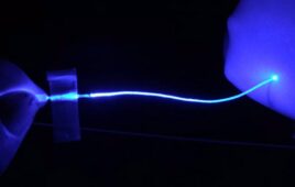
J. Estrada (Franck Lab)/Brown U Still frame filmed at 200,000 frames/sec of a violently collapsing vapor bubble inside a brain-mimicking collagen gel (bubble size is approximately 100 microns). Inside the gel are thousands of brain cells (neurons).
In the fleeting moments after a liquid is subjected to a sudden change in pressure, microscopic bubbles rapidly form and collapse in a process known as cavitation. In mechanical systems such as propellers, the resulting shock waves and jets can cause gradual wear, and in biological systems, they can shred and distort cells. In the human brain, this is believed to be a mechanistic cause of traumatic brain injury, or TBI, but the phenomenon has yet to be directly observed in brain tissue because the bubbles appear and disappear within microseconds.
To better understand the connection between microcavitation and traumatic brain injury, researchers at Brown University have developed a novel 3D imaging system that allows them to film one million frames per second, with a single camera on a single microscope, and ultimately to explore damage to neurons in the laboratory. This will allow them to observe whether cavitation events occur in the tissues surrounding neurons, and if so, it will provide a starting point for identifying the mechanisms of damage in both human and animal tissue samples.
“We still don’t know what the cellular or tissue damage due to primary blast exposure, or blast TBI actually looks like. We also don’t know if it’s due to cavitation or not. This is partially because we don’t know what to look for in MRI, histologies, biopsies and tissue sample scans and images,” said Christian Franck, an assistant professor in Brown University’s School of Engineering.
To address this, Franck and his colleagues are seeking to understand how cavitation might injure neurons by using a 3D imaging system coupled with a diffraction grating to examine their post-exposure morphology. They will present their recent findings at the American Physical Society (APS) Division of Fluid Dynamics (DFD) 68th meeting, to be held Nov. 22-24 in Boston, Mass.
Current 3D image correlation methods typically involve making stereo projections of objects — a geometric mapping function that was used to create the world’s first celestial charts by projecting a sphere onto a plane — by capturing images with two or more cameras. According to Franck, however, this technique doesn’t translate well to observations taken through a microscope, as using multiple cameras carries a loss of spatial resolution. His laboratory focuses on developing experimental techniques to resolve the motion of materials in three dimensions.
In order to capture the three-dimensional motion with a single lens, the researchers placed a diffraction grating in front of the imaging camera on the microscope. They then used a nanosecond-pulsed infrared laser to produce single cavitation bubbles within a model neural network made of collagen and biomimetic hydrogels embedded with neurons, thus simulating the action of a neural network subjected to a negative pressure surge.
When images of the deforming material meet the diffraction grating, they diffract into the first negative, first positive, and zero-order modes. This effectively generates two perspectives of the object — as if you had two cameras — without sacrificing spatial resolution.
“The major challenge for us was to recombine the multiple grating perspectives back onto the same physical location on the camera,” Franck said.
To do this, the researchers spectrally assigned different colors to the -1 and +1 orders that the camera can detect. As all the diffraction orders are initially illuminated with white light, Franck and his colleagues put a red filter in front of where the -1 order light would pass and a blue filter in front of where the +1 light would pass. The camera then registers them as separate RGB colors, which allows the researchers to easily recombine them — thus recreating the 3D motion field at frame rates high enough to observe cavitation.
“This is something really exciting for us, and as far as we know, it has never been done before,” said Franck.
Currently, Franck’s lab is able to resolve motion fields down to the 10-100 nanometer scale. Future work for Franck and his colleagues includes improving their techniques to observe material deformations on the single nanometer level using super-resolution microscopy techniques in 3D.