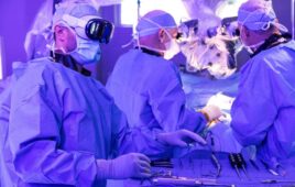
[Image from the University of Alberta]
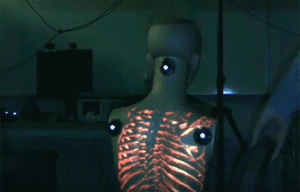
The system ProjectDR projects medical images like CT scans and MRI data directly onto a patient’s body in a way that moves with the body.
“We wanted to create a system that would show clinicians a patient’s internal anatomy within the context of the body,” Ian Watts, a computing science graduate student and the developer of the system, said in a press release.
A motion-tracking system uses infrared cameras and markers on the patient’s body to project the images on a patient’s body using a standard projector. It seems simple, but the challenging part for Watts was having the image track properly on the patient’s body as they moved. As a solution, he developed custom software that connects all of the components.
“There are lots of applications for this technology, including in teaching, physiotherapy, laparoscopic surgery and even in surgical planning,” Watts said.
ProjectDR can project segmented images like if a doctor wants to see only the lungs or only blood vessels.
Watts plans to continue working on the system to improve its automatic calibration and add depth sensors. He also plans to test the program in a clinical setting.
“Soon we’ll deploy ProjectDR in an operating room in a surgical simulation laboratory to test the pros and ons in real-life surgical applications,” Pierre Boulanger, professor in the department of computing science, said.
Watts and the other researchers plan to conduce pilot studies to test the usability of the system for chiropractic and physical therapy procedures. Once the pilot studies are successful, they plan to use the system in real surgical pilot studies.