Back in the day, the survival rate for conjoined twins was low.
Thanks to advanced technology, more have survived and have been successfully separated. So, what’s the next step? Better surgical planning and that’s exactly what 3D technology has allowed surgeons to do. It has allowed surgeons to better plan out various types of surgery; thanks to it, Ximena and Scarlett Hernandez-Torres, twins out of a set of triplets, were given a chance to give separate lives.
A 3D model of the conjoined twins (Credit: 3D Systems, Inc.)
“There’s a 50 million to one chance that triplets will include conjoined twins,” says 3D Systems. “On top of that, there’s a six percent chance that twins of any kind will be joined near the hips.”
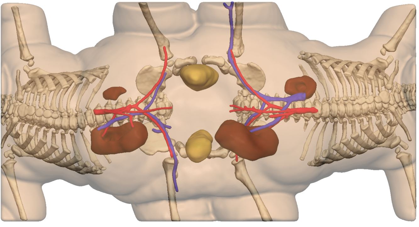
The twins were conjoined from the navel and below, making them share a colon and half of their uterus on each side, according to 3D systems. In addition, their kidneys connected to each other’s bladder.
Prior to the procedure, the doctors spent several months studying the anatomy of the twin, then devised a plan to separate the girls. CT scan data was sent over to the 3D Systems team and they created a 3D digital file of it. From there, 3D-printed anatomical models were developed.
A physicial anatomical models of the conjoined twins, printed with the 3D printer (Credit: 3D Systems, Inc.)
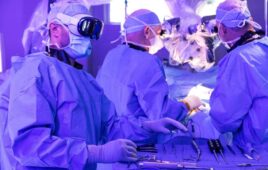
“The physical models were fantastic,” says Dr. Kevin Hopkins. “Unlike a two-dimensional x-ray or 3D visualization, you could hold these models in your hands. They were a great way to show team members exactly where the organs were located, where the cuts would be, and how to position the patient.”
To make these models, the team used a 3D Systems ProX 800 stereolithography (SLA) printer. They also used translucent plastic material for the model that showed the major vasculature and organs involved with the separation. The second model was designed with white plastic material, used to represent the surface of the skin. It allows to better incision planning, as it shows the contours of the skin, Joe Fullerton, Team Lead for Medical Imaging and Modeling at 3D systems points out. Both models are even taken into the operating room for reference during the surgery.
“They are improving all the time,” says Dr. Hopkins. “We expect that both of them will be able to walk and lead a normal life.