The new prosthetic infection detection method out of UC San Diego uses an imaging technique and a thin-film sensor that coats the prostheses, as opposed to having to have an MRI, CAT scan or X-ray to detect infections.
Prosthetic joint infection is a rising problem with arthroplasty surgeries. Most PJIs require surgery and medical therapy to treat the resulting joint pain. The cost to treat PJI in the U.S. was $566M in 2009 and is expected to more than double to $1.62B by 2020, according to a 2014 Clinical Microbiology Reviews article relayed by the National Institutes of Health.
The UC San Diego researchers’ imaging technique is an improved version of an electrical capacitance tomography (ECT) that measures the human tissue and the electrical properties in human tissue and prosthesis.Physicians can measure the data and reconstruct an area’s electrical properties to show the health of the tissue, bone and prosthesis, which is distinguished from the ECT.
Ken Loh, a professor of structural engineering at the Jacobs School of Engineering at UC San Diego, and PhD student Sumit Gupta further improved the algorithm of the ECT by developing a thin-film sensor. The thin-film sensor assists the imaging technique’s ability to detect infections in prostheses used for amputees, knee, hip and other joint replacements.
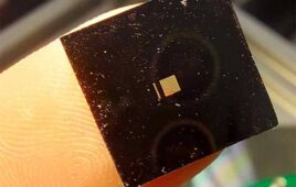
Sensitive to pH, the film is made of a conductive polymer matrix and carbon nanotubes that are inserted in the matrix that increases the prostheses ability to conduct electricity based on pH levels. pH levels are noticeably different when there is an infection in human cells. Fluctuations in pH levels affect the conduciveness of electricity in the ECT.
The thin-film sensor was tested by spray-coating a plastic rod that simulated a prosthetic and manipulating the pH levels. The imaging technique then scanned the plastic rod and measured the electrical properties for the ECT.
[Want to stay more on top of MDO content? Subscribe to our weekly e-newsletter.]


