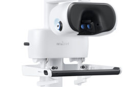
Surgical Information Sciences (SIS) recently won FDA clearance for software designed to guide deep-brain stimulation (DBS) procedures.
The Plymouth, Minn.-based company’s software uses deep learning models to identify brain structures such as the subthalamic nuclei (STN) and globus pallidus internus/externus (GPi/GPe) commonly targeted for DBS procedures. The SIS system provides visualization of these structures for surgical planning and for location of the implanted DBS leads and lead contacts post-surgery.
The STN and GPi/GPe are key targets for patients with Parkinson’s disease, but are too small to be accurately visualized with today’s imaging capabilities, according to SIS. SIS designed its technology to produce 3D anatomical models of brains, uniquely defined for each patient, created by using proprietary innovations in high–field MRI (7 Tesla or “7T”) acquisition combined with proprietary data processing and analysis algorithms. The company claims its software generates patient-specific images of the STN and GPi/GPe with approximately 70% better accuracy than the atlas-based systems used today.
“Placing a lead accurately in the target can make all the difference in patient outcomes and symptoms after DBS surgery” said Dr. Kyle Nelson, neurosurgeon from Metropolitan Neurosurgery in Minneapolis, who conducted the first commercial use with the software. “After comparing SIS to my standard target planning and intra-operative testing, I found the SIS image to be valid. SIS is more accurate than standard surgical planning programs I’ve used, and leaves no doubt as to where the target structure is located.”
The FDA granted updated clearance for the software in April.
“This marks a significant milestone for SIS as we have spent nine years building the most accurate visualization tool for the key targets used in DBS procedures for the treatment of Parkinson’s. To finally have it used is very satisfying,” said SIS president & CEO Brad Swatfager in a news release. “The improved accuracy of the visualized structure coupled with the ease of image use in the planning or programming should significantly improve the experience for physicians and patients.”
![A photo of the Medtronic GI Genius ColonPro polyp detection system flagging a potential sign of colon cancer during a colonoscopy. [Photo courtesy of Medtronic]](https://www.medicaldesignandoutsourcing.com/wp-content/uploads/2024/04/Medtronic-GI-Genius-doctors-268x170.jpg)