On the left, in vivo image of nanomachines using current microscopy techniques; on the right, the new method allows 3-D observation of nanomachines in vivo and provides 25-fold improvement in precision (Credit: O. Gallego, IRB Barcelona)
Currently, biologists who study the function of protein nanomachines isolate these complexes outside the cell in test tubes, and then apply in vitro techniques that allow them to observe their structure down to the atomic level. Alternatively, they use techniques that allow the analysis of these complexes within the living cell but that give little structural information.
In this study, the scientists have directly observed the structure and function of the protein machinery in living cells.
“The in vitro techniques available are excellent and allow us to make observations at the atomic level, but the information provided is limited. We will not know how an engine works if we dissemble it and only look at the individual parts. We need to see the engine assembled in the car and running. In biology, we still do not have the tools to observe the inner workings of a living cell, but the technique that we have developed is a step in the right direction and we can now see, in 3D, how the protein complexes carry out their functions,” explains Oriol Gallego, IRB Barcelona researcher and coordinator of the group that undertook this study, which also involved PhD student Irene Pazos.
Watching the nanometric machinery at work
The new strategy uses methods from super-resolution microscopy and computational modelling.
The technology allows researchers to observe protein complexes with a precision of 5 nm, a resolution “four times better than that offered by super-resolution and that allows us to perform cell biology studies that were previously unfeasible,” explains Gallego.
The researchers genetically modify cells in order to build artificial supports inside onto which they can anchor protein complexes. These supports are designed in such a way as to allow them to regulate the angle from which the immobilized nanomachinery is viewed. Then, in order to determine the 3D structure of the protein complex, they use super-resolution techniques to measure the distances between different components and then integrate them in a process similar to that used by GPS.
Basic features of exocytosis
Gallego has used this method to study exocytosis, a mechanism that the cell uses to communicate with the cell exterior.
For instance, neurons communicate with each other by releasing neurotransmitters via exocytosis. The study allows the scientists to reveal the entire structure of a key nanomachine in exocytosis that until now was enigmatic.
“We now know how this machinery, which is formed by eight proteins, works, and what each protein is important for. This knowledge will help us to better understand the involvement of exocytosis in cancer and metastasis—processes in which this nanomachinery is altered,” he explains.
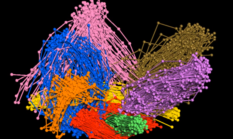
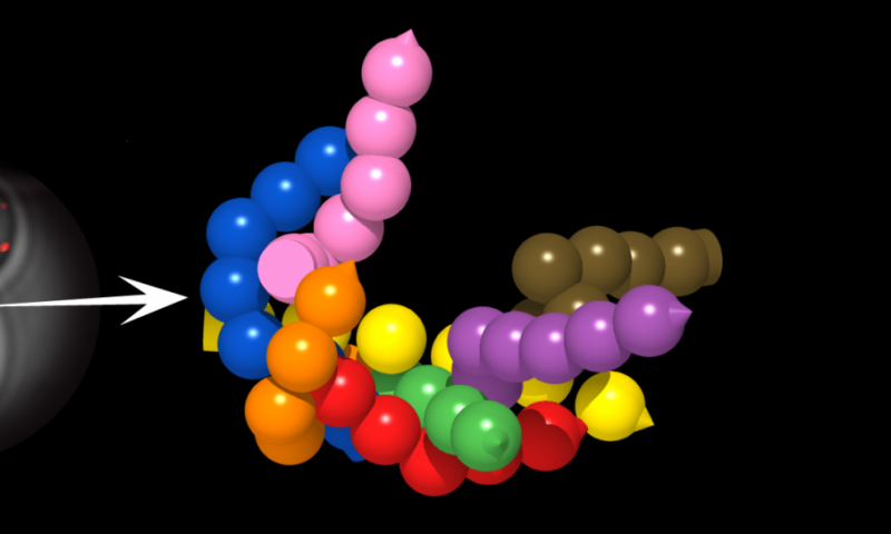
A model of the architecture of the main machinery involved in exocytosis. The eight proteins, each shown in a different color, are bound, forming the nanomachine or protein complex. (Credit: O. Gallego, IRB Barcelona)
An understanding of how nanomachines carry out their cell functions has biomedical implications since alterations in the inner workings can lead to the development of diseases.
With this new strategy, it will be possible to study cellular protein machinery in health and in disease. For example, it would be possible to see how viruses and bacteria use protein nanomachines during infection, and to better understand the defects in complexes that lead to diseases in order to design new therapeutic strategies that reverse them.
The technique can be used on relatively large complexes.
“Being able to see protein complexes measuring 5 nm is a great achievement, but there is still a long way to go toward observing the inside of the cell at the atomic scale that in vitro techniques would allow,” says Gallego. “But I think that the future lies in integrating various methods and combining the power of each one.”


