Medical Design and Outsourcing

  • Home
  • Medical Device Business
    • Mergers & Acquisitions
    • Financial
    • Regulatory
  • Applications
    • Cardiovascular
    • Devices
    • Imaging
    • Implantables
    • Medical Equipment
    • Orthopedic
    • Surgical
  • Technologies
    • Supplies and Components Index
    • Contract Manufacturing
    • Components
    • Electronics
    • Extrusions
    • Materials
    • Motion Control
    • Prototyping
    • Pumps
    • Tubing
  • MedTech Resources
    • Medtech Events in 2025
    • The 2024 Medtech Big 100
    • Medical Device Handbook
    • MedTech 100 Index
    • Subscribe to Print Magazine
    • DeviceTalks
    • Digital Editions
    • eBooks
    • Manufacturer Search
    • Podcasts
    • Print Subscription
    • Webinars / Digital Events
    • Whitepapers
    • Voices
    • Video
  • 2025 Leadership
    • 2024 Winners
    • 2023 Winners
    • 2022 Winners
    • 2021 Winners
  • Women in Medtech
  • Advertise
  • Subscribe

Fluorescent Holography: Upending The World Of Biological Imaging

October 25, 2016 By Colorado State University

Optical microscopy experts at Colorado State University are once again pushing the envelope of biological imaging.

Jeffrey Field, a research scientist in electrical engineering and director of CSU’s Microscope Imaging Network, has designed and built a fluorescence-detection microscope that combines three-dimensional and high-resolution image processing that’s also faster than comparable techniques.

The work, with co-authorship by Randy Bartels, professor of electrical and computer engineering, and former postdoctoral researcher David Winters, has been published in Optica, the journal of the Optical Society of America. They named their new microscope CHIRPT: Coherent Holographic Image Reconstruction by Phase Transfer.

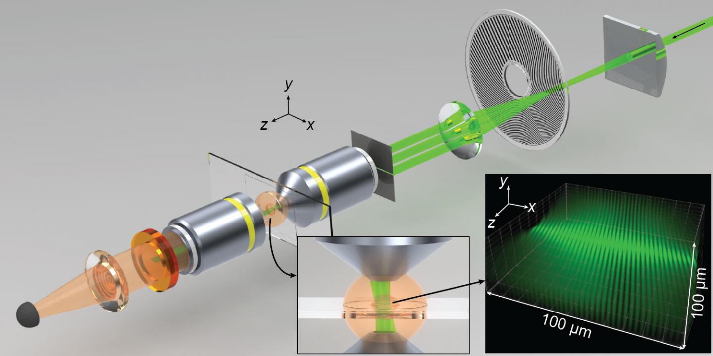
Spatiotemporal modulations of illumination intensity in the CHIRPT microscope are achieved by imaging a spinning modulation mask to the focal plane of the microscope. A spatial filter placed in the pupil plane of the objective lens allows illumination intensity to form by the interference of two beams in the object plane. The microscope and illumination intensity are shown here at a snapshot in time. (Credit: Jeff Field/Colorado State University).

Field and other optics scientists work in a world of tradeoffs. For example: an advanced deep-tissue imaging technique called multiphoton fluorescence microscopy employs a short, bright laser pulse focused tight to one spot, and the fluorescence intensity from that one spot is recorded. Then, the laser moves to the next spot, then the next, to build up high-resolution 3D images. The technique offers subcellular detail, but it’s relatively slow because it illuminates only one tiny spot at a time.

Other techniques, like spinning disk confocal microscopy, are faster because they shine light on multiple spots, not just one, and they scan simultaneously over a larger area. But unlike multiphoton, these techniques require collecting an image with a camera. As a result, fluorescent light emitted from the specimen is blurred on the camera, leading to loss in resolution, and with it, subcellular detail.

Call them greedy, but Field and colleagues want it all.

Their goal is working around each of these limitations – speed, resolution, size of field – to break through established boundaries in light microscopy.

Field and Bartels’ new microscope builds upon a previously published technique, and permits digital re-focus of fluorescent light. It illuminates not one point, but multiple points by harnessing delocalized illumination spread over a large area. The physical principles they are using are similar to holography, in which scattered light is used to build a 3-D image.

Using a large illumination field, followed by back-end signal processing, the microscope can define distinct light modulation patterns of many points within the field of view. It builds up a 3-D image by combining the signals from all those distinct patterns.

“The idea is that you have a fluorophore at any point in the specimen, and the temporal structure of its fluorescence will be distinguishable from all others,” Field said. “So you can have this huge array of fluorophores, and just with this single-pixel detector, you can tell where every one of them is in that 2D field.”

So what does this new technique allow? Deep-tissue images in three dimensions, with better depth of field than comparable techniques. Depth of field, like in photography, means background images are in sharp focus along with the main image. And the CSU researchers can work at 600 frames per second, which is many times faster than established techniques.

With their new microscope, images can also be post-processed to remove aberrations that obscure the object of interest. It’s akin to being able to focus a picture after it’s been taken.

The CHIRPT microscope could allow biomedical researchers to produce sharp, 3-D images of cells or tissue over a much larger volume than conventional fluorescence microscopy methods allow. It could lead to things like imaging multicellular processes in real time that, with a conventional light microscope, could only be seen one cell at a time.

Related Articles Read More >

A photo of nitinol, a nickel-titanium alloy used for medical devices such as stents, heart valves, catheters and orthopedics.
What is nitinol and where is it used?
An image of Abbott's Infinity deep brain stimulation (DBS) implants and leads.
How Abbott developed the first-of-its-kind Infinity DBS system
Axoft Fleuron brain-computer interface BCI probe
Axoft makes Fleuron BCI material available for purchase, inks license deal with Stanford
An illustration showing the Edwards Lifesciences Sapien M3 transcatheter mitral valve replacement (TMVR) system's valve being placed in the heart. [Image courtesy of Edwards Lifesciences]
The top nitinol cardiac medtech news of 2025 (so far)
“mdo
EXPAND YOUR KNOWLEDGE AND STAY CONNECTED
Get the latest medical device business news, application and technology trends.

DeviceTalks Weekly

See More >

MDO Digital Edition

Digital Edition

Subscribe to Medical Design & Outsourcing. Bookmark, share and interact with the leading medical design engineering magazine today.

MEDTECH 100 INDEX

Medtech 100 logo
Market Summary > Current Price
The MedTech 100 is a financial index calculated using the BIG100 companies covered in Medical Design and Outsourcing.
DeviceTalks

DeviceTalks is a conversation among medical technology leaders. It's events, podcasts, webinars and one-on-one exchanges of ideas & insights.

DeviceTalks

New MedTech Resource

Medical Tubing

MassDevice

Mass Device

The Medical Device Business Journal. MassDevice is the leading medical device news business journal telling the stories of the devices that save lives.

Visit Website
MDO ad
Medical Design and Outsourcing
  • MassDevice
  • DeviceTalks
  • MedTech100 Index
  • Medical Tubing + Extrusion
  • Medical Design Sourcing
  • Drug Delivery Business News
  • Drug Discovery & Development
  • Pharmaceutical Processing World
  • R&D World
  • About Us/Contact
  • Advertise With Us
  • Subscribe to Print Magazine
  • Subscribe to our E-Newsletter
  • Listen to our Weekly Podcasts
  • Join our DeviceTalks Tuesdays Discussion

Copyright © 2025 WTWH Media, LLC. All Rights Reserved. The material on this site may not be reproduced, distributed, transmitted, cached or otherwise used, except with the prior written permission of WTWH Media LLC. Site Map | Privacy Policy | RSS

Search Medical Design & Outsourcing

  • Home
  • Medical Device Business
    • Mergers & Acquisitions
    • Financial
    • Regulatory
  • Applications
    • Cardiovascular
    • Devices
    • Imaging
    • Implantables
    • Medical Equipment
    • Orthopedic
    • Surgical
  • Technologies
    • Supplies and Components Index
    • Contract Manufacturing
    • Components
    • Electronics
    • Extrusions
    • Materials
    • Motion Control
    • Prototyping
    • Pumps
    • Tubing
  • MedTech Resources
    • Medtech Events in 2025
    • The 2024 Medtech Big 100
    • Medical Device Handbook
    • MedTech 100 Index
    • Subscribe to Print Magazine
    • DeviceTalks
    • Digital Editions
    • eBooks
    • Manufacturer Search
    • Podcasts
    • Print Subscription
    • Webinars / Digital Events
    • Whitepapers
    • Voices
    • Video
  • 2025 Leadership
    • 2024 Winners
    • 2023 Winners
    • 2022 Winners
    • 2021 Winners
  • Women in Medtech
  • Advertise
  • Subscribe