[Image from GE Healthcare]
Denis Ducreux has been fascinated by the brain since he was a child. He always wanted to understand its functions and possibilities better.
“I am amazed by the brain’s beauty, organization, structure, reflecting the universe’s organization,” Ducreux, who is also the head of the diagnostic neuroradiology department at Bicêtre Hospital in France, said in a press release. “The brain is the center of all knowledge, emotions, memories, behavior and creations.”
While studying radiology, physics and neurosciences, Decreux became intrigued by connections and white matter fibers that create the limbic bundles in the brain. The limbic bundles are responsible for emotion, behavior, motivation, long-term memory and sense of smell.
“Limbic bundles are the ‘unconscious’ or ‘instinctive’ freeways of the brain, related to memories, behavior and emotions. That’s why all my images are focusing on the limbic system because it’s the center of the unconscious,” Ducreux said in a press release.
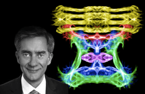
Ducreux uses GE Healthcare’s 3T Signa Architect MRI system to capture raw MRI images, then uses a research-use-only neuroimaging software, known as BrainAnalyst, to track the fibers in the brain. He has used the process since 2017 but has been creating renderings of brain fibers using MRI images for 15 years.
“Signs Architect gave me the possibility to see tiny details in the brain, which enlightened all the images I created. Other MRI systems failed to do such an impressive imaging of the white matter fibers,” Ducreux said.
Ducreux uses a 48 channel head coil to capture clear MRI images and a specific diffusion tensor imaging (DTI) sequence that he created. The DTI sequence is an MRI-based neuroimaging technique that allows him to estimate the location, orientation and anisotropy of the white matter tracts in the brain.
“I can see tiny sub-millimeter details, which in the limbic system are especially of great interest to me,” Ducreux said.
Ducreux says that the images he captures from the Signa Architect could be used in research to better understand how humans’s emotional processing works.
“The limbic system refers to man’s inner temple, the ‘unconscious,’ where behavior, emotions, memory are processed,” Ducreux said. “With MRI images, you can track fibers in the brain, which gives another view of the structures in the brain as well as the functions of the brain. For the physician, it could potentially help them identify which part of the brain is damaged or functioning normally.”
He also suggests that high-resolution imaging from the MRI system could help doctors and researchers understand other complex disorders like addiction and mental illness. Ducreux is currently doing research on how to use artificial intelligence to diagnose neurological diseases, as well as studying difference neurological processes.
“When you’re analyzing the unconscious, you have wider access to the brain and your behavior. So what we can learn is how to control ourselves or to know why we have this kind of behavior in mind, and when you control the unconscious, you also can control the addictions,” Ducreux said. “I want to promote the power of our unconscious and the power of our emotions, and beneath all these, to promote the power of our unconscious may always fool ourselves, and we have to be aware of that to behave appropriately.