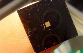
These 1.5 mm capsules are embedded with a fluorine-containing compound that allows the researchers to monitor their oxygen levels with MRI once implanted in the body. [Image from MIT]
The researchers suggest that the pancreatic islet cells could monitor blood glucose and secrete insulin as needed. However, the implanted cells need to receive enough oxygen to produce insulin in order for the transplantation to be successful. The MIT researchers have developed a way to measure oxygen levels in the cells over a set period of time in living animals to predict which pancreatic islet cell implants would be effective.
A specialized type of magnetic resonance imaging was able to track how oxygen levels of implanted cells in the intraperitoneal (IP) cavity could move through the cavity over time. The method was tested in mice.
“Our goal is to make living cellular factories that can supply drugs on demand for patients. The ability to track the oxygen supply and the location of implanted cells will help us better understand how to build and use successful therapies,” Daniel Anderson, an associate professor in the department of chemical engineer and the senior author on the study, said in a press release.
Anderson and his team have been developing implantable islet cells encapsulated in alginate particles for several years. Alginate particles could replace pancreatic islet cells in people who have Type 1 diabetes.
In a study, the researchers discovered that larger particles with a diameter of 1.5 mm are able to function longer than smaller, 0.5 mm diameter particles. The smaller particles typically end up surrounded by scar tissue, blocking access to oxygen.
Particles move through the IP once implanted, which means their oxygen exposure needs to be tracked. The IP space has varying levels of oxygen. Other studies have shown that smaller particles cluster in patches of fat, which typically have less oxygen, making the particles fail.
The MIT researchers used a fluorine MRI to measure oxygen levels in living tissue. Fluorine MRIs measure interactions between a magnetic field and fluorine nuclei and how the interactions are affected by oxygen.
In the study, the researchers used a fluorine-containing material called perfluorocarbon emulsion into the alginate that they had used to encapsulate the islet cells.
Particles with diameters of 0.5 and 1.5 mm were tested in both diabetic and non-diabetic mice. Non-diabetic mice received alginate implants with no cells inside. Diabetic mice received implants with pancreatic islet cells. The fluorine MRI measured the oxygen levels in the IP space for three months. The researchers also measured the diabetic mice’s blood glucose levels.
The researchers used a machine-learning algorithm to analyze the resulting data and go through the images to find correlations between the position of the capsules in the IP space, the oxygen levels and blood glucose levels of the mice.
“These kinds of imaging studies involve a lot of data, and screening all of these 2D images and making decisions about how the position of the capsules affects oxygen concentration is extremely challenging and very error prone when it’s done by a human observer,” Virginia Spanoudaki, the lead author on the study, said. “So we relied on machine learning to automatically go through the images and find associations between the positions of the capsules and other parameters.”
The data showed that smaller capsules could produce enough insulin to treat diabetic mice in the first 30 days of treatment, but then start to gather in large clusters in fatty areas of the animal’s extremities, according to the researchers. The larger capsules spread out over a larger area and migrated to both high- and low-oxygen areas. The cells from the larger capsules could also secrete enough insulin to keep blood glucose levels stable over several months.
“The MIT group has previously shown the better transplant results in mice (and non-human primates) using capsules with a diameter of 1.5 mm compared with 0.5 mm,” said Gordon Weir, the co-head of the Joslin Diabetes Center’s section on islet and regenerative biology, said. “Now with this remarkable technique, we can see what we suspected: that the smaller capsules tend to clump more easily, which results in a more hypoxic environment that leads to impaired insulin secretion and more cell death.”
Anderson and his research team started a company called Sigilon Therapeutics to further develop the bioartificial pancreas. They hope to begin testing implantable islet cells in patients by early 2020. They also hope to implement the oxygen measurement technique in larger animals like humans to develop different versions of the encapsulated islets.
“Based on measurements in larger animals, we would like to understand whether there are different ways to design the bioartificial pancreas so that this aggregation of capsules that potentially results in reduced oxygen does not happen,” Spanoudaki said. “We are hoping to use this as a guide to make better designs for the bioartificial pancreas.
The research was published in the Proceedings of the National Academy of Sciences and was supported by JDRF, the Leona M. and Harry B. Helmsley Charitable Trust Foundation, the Parviz Tayebati Research Fund and a Koch Institute Support Grant from the National Cancer Institute.