[Image from unsplash.com]
Combining squid ink with light and ultrasound, engineers at UCSD have created a new dental imaging method that can painlessly and non-invasively examine a patient’s gums. The method is also more comprehensive and more accurate than current methods.
“The last time I was at the dentist, I realized that the tools that are currently being used to image teeth and gums could use significant updating,” Jesse Jokerst, a nano engineering professor at UCSD and senior author on the study, said in a press release.
Currently, dentists use an instrument called the periodontal probe to determine gum health. The probe is a thin, hook-like metal tool that is inserted between the teeth and gums to determine how far the patient’s gums have shrunk back from the teeth and created pockets. Pocket depths that are one to two millimeters shows healthy gums. Three millimeters or more could be an indicator of gum disease.
Gum disease, if gone untreated, can cause tooth loss and other health problems. A 2010 Centers for Disease Control study found that almost half of adults over 30 in the U.S. have some form of a periodontal disease.
The periodontal probe is invasive and can sometimes cause discomfort with a patient. Measurements also differ between dentists and the probe can only measure pocket depth of one spot at a time. The squid ink method, however, can reduce the pain and image an entire pocket depth around teeth consistently and accurately.
“Using the periodontal probe is like examining a dark room with just a flashlight and you can only see one area at a time. With our method, it’s like flipping on all the light stitches so you can see the entire room all at once,” Jokerst said.
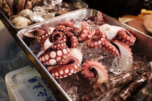
To use the squid ink method, the patient first has to rinse their mouth using a paste made of commercially-available food-grade squid ink mixed with water and cornstarch. The squid ink acts as a contrast agent for a photoacoustic ultrasound. A photoacoustic ultrasound shines a light signal like a short laser pulse onto a sample that heats up and expands, creating an acoustic signal that can be analyzed.
During the rinsing, the squid ink’s natural melanin nanoparticles absorb light and get trapped in the pockets. Researchers can then shine a laser light onto the area where the squid ink will heat up and swell quickly which creates pressure difference in the pockets that ultrasounds can detect. The ultrasound allows researchers to create a full map of pocket depths around each tooth.
The UCSD researchers tested the photoacoustic method in a pig model that had a mix of shallow and deep gum pockets. The results were similar to the ones they got using a periodontal probe and were consistent across several tests. Periodontal probe measurements varied widely among different tests.
“It’s remarkable how reproducible this technique is compared to the gold standard,” Jokerst said.
The team hopes to collaborate with dentists in the future and test their method in humans. They also hope to refine the squid ink so that the taste is more tolerable and less salty and bitter. The researchers also want to replace laster lights with inexpensive and portable light systems like LEDs and develop a mouthpiece that has the technology in it to measure periodontal health.
The research was funded by the National Institutes of Health through grants and was published in the Journal of Dental Research.

Wow its a very interesting post. Squid ink is going to be very effective technique. Thanks for sharing…