Clinical doctors and laboratory scientists, working together in the same labs, are conducting research into issues surrounding oral cancer detection. Wanting to aid surveillance and early tumor diagnosis, the Biophotonics Research Group at King’s College London’s Dental Institute have developed a new clinical imaging instrument that will potentially reduce the number of biopsies required in the future.
Oral or mouth cancer is where a lesion develops in the lining surface of the tongue, mouth, lips and gums. Diagnosis is difficult as many other safer conditions can resemble the early stages of the disease, and several can transform into cancers at random points.The traditional stereotype of an oral cancer patient as a hard drinking and smoking individual is also now evolving towards younger non-smokers and drinkers. Such patients are even harder to spot as their lesions are developing without the traditional risk factors being present, and can be more aggressive too.
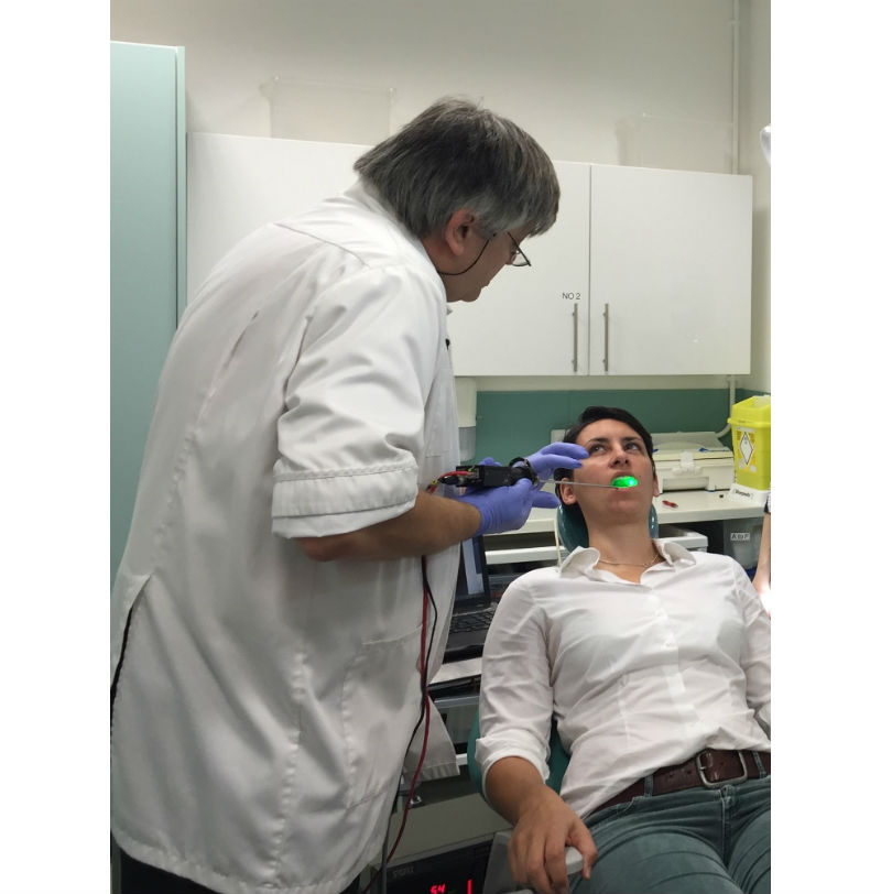
The prototype, named the Microvascular scope is a simple, portable and functional device. (Credit: King’s College London)
Biopsy is the current “gold standard” for characterizing abnormal tissue from the mouth. It is an invasive and painful procedure where suspected cancerous tissue is removed and tested, and in itself is often a deterrent for getting a lesion checked by an oral health practitioner.
Over the last 40 years, despite many technique and approach developments, the five-year survival figures of approximately 50 percent remain resolutely static. Many authorities feel that late presentation and diagnosis remains a key determinant in final outcome.
The Device
The cost-effective and non-invasive approach developed by the researchers has already identified the location of invasive cancers as small as 1mm in diameter, allowing curative minimally invasive surgery with fewest residual scars and deformities.
The prototype, named the Microvascular scope is a simple, portable and functional device that could change the way clinicians view oral cancer detection by allowing the host’s first response to the tumor – new blood vessel growth (angiogenesis) – to be imaged undisturbed and as it occurs, at extremely high resolution.
“A device that can help survey suspicious lesions and guide biopsies to more accurately confirm the existence of cancer and its exact location would prove invaluable,” Dr. Richard Cook from the dental institute explained. “Biopsy is an elaborate and time-consuming process and is unpleasant, deterring patients from further treatment. There is often the need for biopsies in multiple locations to ensure all areas of a potentially cancer bearing lesions are correctly assessed. This is often repeated over time to rule out misdiagnosis.”
In addition to Cook, the Biophotonics research group includes Dr. Frederic Festy, professor Timothy Watson and Dr. Neveen Hosny, along with a diverse group of clinicians, engineers, physicists, and bioengineers. The functional prototype is ready for commercialization, according to the researchers.
Mouth cancer occurs when something goes wrong with the normal cell lifecycle, causing them to grow and reproduce uncontrollably. Smoking, drinking alcohol and infection with the human papilloma virus (HPV), the virus that causes genital warts, can all increase the risk of developing mouth cancer.