This article will appear in the upcoming May print issue of Surgical Products.
 The many refinements in breast conservation therapy have resulted in greatly improved treatment. But surgeons and radiation oncologists still face challenges in working together to more accurately target radiotherapy that is both clinically effective and achieves the best cosmetic result.
The problem isn’t the radiotherapy delivery methods themselves. Technologies such as 3D conformal, intensity modulated radiotherapy/image-guided radiotherapy, brachytherapy, volumetric modulated arc therapy, and multi-beam approaches are admirably precise. But, until this time, they have not been widely used in the treatment of breast cancer.
The reason these methods are not more commonly used for breast cancer patients is the lack of a reliable way to delineate the target area with a high degree of accuracy. Without that, treatment planners can’t always take advantage of the technology at their command.
But now, this too appears to be changing for the better. Recently, a new approach has emerged that marks the target area with greater, reproducible accuracy and it appears to have other advantages for post-operative radiotherapy as well. This approach uses a new medical device that is placed by a surgeon when tissue is removed. The surgically implantable, spiral incorporates six permanent titanium clips that are “suspended” in a fixed three-dimensional arrangement that can clearly be identified.
The marker is sutured to the lumpectomy cavity at the time of tumor excision after removal of the tumor and surrounding margins is complete. The spiral is made of bioabsorbable material that is reabsorbed by the patient’s body slowly over time. The titanium clips remain, because they can provide specific, stable, long-lasting landmarks so the site of the excised tumor can be clearly visualized over time.
To understand how best to use this three-dimensional tissue marker (called BioZorb, made by Focal Therapeutics), it helps to review the current state of how physicians identify the lumpectomy site within the breast after tissue is removed. The surgical area can be identified by using small surgical clips individually placed during surgery, or physicians can rely on external anatomic landmarks and/or tissue changes surrounding the seroma cavity as seen on a CT scan. Each of these current methods can be somewhat problematic. For instance, the clips can migrate during the post-surgical healing process or the seroma cavity may be gone by the time radiation treatments are delivered, thus leaving the surgical site unidentifiable within the breast.
This means there’s often guesswork in defining the area needing therapy. To compensate, treatment planners must err on the side of caution to prevent a cancer recurrence. In other words, they typically can target a much larger area than necessary.
The result: adjacent healthy tissues can be unintentionally damaged by exposure to radiation when treating the cancer site. This may negatively affect the cosmetic outcome and, more importantly, can also lead to complications such as harm to nearby structures like the heart or lungs.
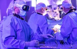
In my surgical practice, I have implanted this 3D marker in more than 60 patients. Its performance has been impressive and virtually trouble-free. I have not seen any device-related complications – nor have I needed to remove any devices because of an infection or other issue. Also, patient acceptance of the new marker is very high. The cosmetic results have been excellent because the 3D marker allows treatment planners to target the radiation much more tightly than other markers. This minimizes exposure to healthy tissue and bodily structures. The device is placed using standard surgical techniques.
For the marker to be more widely used, of course, its usefulness has to be confirmed by other surgeons and physicians as well. Several centers have presented promising results, and four of my colleagues and I have presented our results at the 2013 annual meeting of the American Society of Breast Surgeons.
We reported on our initial series of patients, which involved 15 women who were implanted with the device. Most of them received standard whole breast irradiation (WBI) and were given a boost to the tumor bed. In five patients we were able to compare boost volumes with and without the 3D marker. When the 3D marker was used, we typically (4 of 5) saw a 30 percent volume reduction in the boost volume, compared to traditional methods (mean reduction 59 percent).
Another intriguing finding concerned the seroma. The tissue changes around the seroma cavity are often used as a marker for radiotherapy. In our patients, however, six of the 15 (40%) had no seroma or fluid outside the area outlined by the tissue marker. The 3D marker was able to identify the site in some of those cases — meaning it was useful in instances where traditional methods could not be employed.
The oncology team (including the physician and physicist) rated their experience. The marker was rated “easily visible” in all 15 cases. The team also rated the device “very useful” in assisting with boost dose planning in 12 out of 15 (80%) patients. For two of the three remaining patients, the team rated it “fairly useful.” It was not used for treatment planning in one case because circumstances required treatment beyond the zone outlined by the marker.
Yet another important finding concerned so-called respiratory motion, which can introduce additional uncertainty in delivering radiotherapy. This occurs when the patient’s breast moves during breathing, making it difficult to track the target for radiation. Our pilot series found the 3D marker enabled image-based tracking of the lumpectomy cavity (as opposed to a surrogate target) during respiratory motion. This allowed us to use the marker to assist with image-guided radiation delivery. We also found that the marker assisted with patient positioning and setup between each daily radiotherapy fraction, offering a new method of standardized consistency in positioning, and improving the efficiency of setting up for treatment delivery.
The other centers using the device regularly have reached similar conclusions. It’s true that our experience with this new marker is still fairly early, and based on small numbers of patients. But our results are consistent with good experiences doctors are having both in the U.S. and also in New Zealand, where several surgeons have placed the device.
This novel marker appears to solve a targeting issue that has hindered post-lumpectomy radiotherapy, while being easy for surgeons to place. Unlike older methods, it is true to the surgical cavity location every time – because it is directly sutured into the site at the time of tissue removal and it is easy for treatment planners to see using standard imaging. That translates to substantially reduced planning treatment volumes compared to older methods, meaning a chance for shorter length of treatment, and hopefully fewer complications and better results.
Breast conservation therapy was created to produce these kinds of results, but has been held back by the targeting problem. This new 3D marker looks like it can help breast conservation achieve its original aim of treating the cancer while preserving the breast.
To subscribe to Surgical Products magazine, click here.