Hitachi High-Tech and RIKEN, one of Japan’s national institutes for scientific research, announced a jointly developed “MirrorCLEM,” a system for simplifying correlative light and electron microscopy (CLEM) which enables the observation of one using both light and scanning electron microscopes (SEM).
Various types of microscopes are used in a wide variety of fields such as nanotechnology, materials, medical and life sciences. In the medical and life sciences field in particular, SEMs are used to clarify the ultrastructure of cells and tissues, while a type of light microscope called fluorescence microscopes are being used increasingly to observe the localization and behavior of proteins at the molecular level.
In recent years, CLEM techniques, which correlate electron microscopy with fluorescence microscopy, have been developed. However, the observation of the same field in a specimen with microscopes at different magnifications and observation characteristics remained a difficult task.
To solve this problem, Hitachi High-Tech worked with a group led by Dr. Kiminori Toyooka, a senior research scientist with the RIKEN Center for Sustainable Resource Science, to research and develop a system for observing the ultrastructure of organelles containing green fluorescent proteins (GFP).
Overview of the MirrorCLEM System. (Credit: Business Wire)
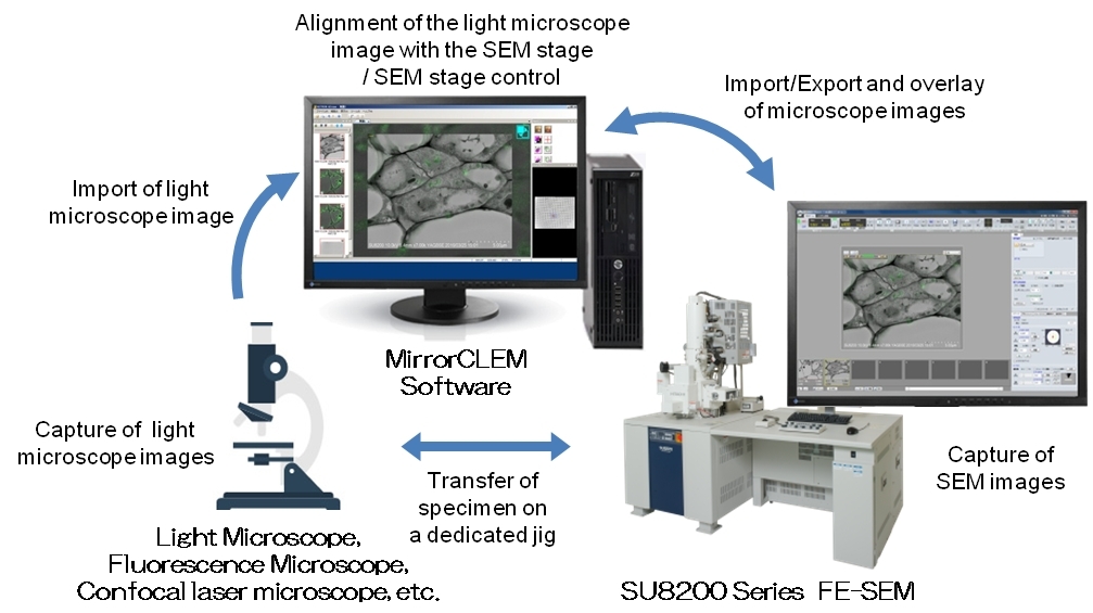
RIKEN developed the microscopic observation workflow and the preparation method of embedding in resin to preserve both the GFP fluorescence and ultrastructure. Meanwhile, Hitachi High-Tech developed a dedicated jig for observing plastic sections mounted on cover slips under a Field-Emission SEM (FE-SEM), as well as the software for swiftly and accurately observing the same location of florescence microscopy in a FE-SEM.
The MirrorCLEM system that Hitachi High-Tech and RIKEN subsequently developed supports quick and accurate CLEM analysis by using a FE-SEM. With this system, the plastic sections can be observed under a light microscope from low magnifications to magnifications that are high enough to clearly observe the structure of interest.
In addition, the FE-SEM stage can be coordinated to the target position in the low-magnification (LM) image, which is observed with the light microscope. Subsequently, the field of view (FOV) in a FE-SEM can move to any region of interest in the LM image and the same FOV can be observed in the FE-SEM under the MirrorCLEM system. Also, the system is capable of displaying an overlay of the light microscope and FE-SEM images in real time. The Hitachi SU8220 FE-SEM equipped with the MirrorCLEM system has already been used to clarify the ultrastructure of peroxisomes containing GFP in the cotyledon and root in transgenic Arabidopsis thaliana.
MirrorCLEM will be launched for sale by Hitachi High-Tech on July 25th, 2016.