[Image courtesy of Anaïs Leproux, Beckman Laser Institute]
“We use visible and near-infrared light at very low power and project it onto the breast. We are trying to characterize the skin damage during radiation therapy, especially for the treatment of breast cancer,” said Anaïs Leproux, a Beckman Laser Institute post-doctoral researcher lead author of the paper on the research. Leproux will present on the work at the OSA Biophotonics Congress: Optics in the Life Sciences meeting, April 2–5 in San Diego.
The research matters because there is presently no method to predict the severity of the late effects of breast cancer radiation treatment, according to The Optical Society. Beyond the skin irritation, peeling and blistering experienced by all women undergoing the therapy, later side effects include permanent skin discoloration and breast tissue thickening.
Leproux and her colleagues are seeking to use the near-infrared light imaging to produce precise measurements of tissue skin toxicity throughout radiation treatment. The hope is to better understand the factors leading to acute and late side effects–and predict them using the imaging technique.
“The toxicity is basically the skin damage, a side effect from the radiation,” Leproux said. “There are a wide range of side effects that we’re observing; erythema [skin redenning], hyperpigmentation, discoloration, dry or wet desquamation [peeling]. Necrosis can happen but is less common.”
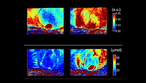
During the imaging, 8 different wavelengths of visible and near-infrared light from LEDs scatters on the skin, with some absorption, according to Leproux. “The reflected light is detected by a camera. Basically, you’re measuring the absorption and the scattering properties of the tissue,” Leproux said.
Spatial Frequency Domain Imaging, or SFDI, has light from the LEDs spatially modulated, with a digital micro-mirror device within the instrument imparting distinct patterns. By measuring how much of each wavelength is absorbed by the skin, Leproux and her colleagues can come up with quantitative values that indicate skin health.
“Since we use several wavelengths of light, we perform spectroscopy and obtain the content of melanin, tissue hemoglobin, in the de-oxygenated and oxygenated state, from which we can calculate the total blood volume and oxygen saturation in the tissue,” Leproux said. “We measure superficially, about 3 to 5 mm deep.”
“We’re hoping that we can see skin thickening in the scattering parameters we’re looking at. We think that the radiation induces a remodeling of the collagen in the skin, which should be seen as a change in the scattering parameter,” Leproux added.
[Want to stay more on top of MDO content? Subscribe to our weekly e-newsletter.]