Medical Design and Outsourcing

  • Home
  • Medical Device Business
    • Mergers & Acquisitions
    • Financial
    • Regulatory
  • Applications
    • Cardiovascular
    • Devices
    • Imaging
    • Implantables
    • Medical Equipment
    • Orthopedic
    • Surgical
  • Technologies
    • Supplies and Components Index
    • Contract Manufacturing
    • Components
    • Electronics
    • Extrusions
    • Materials
    • Motion Control
    • Prototyping
    • Pumps
    • Tubing
  • MedTech Resources
    • Medtech Events in 2025
    • The 2024 Medtech Big 100
    • Medical Device Handbook
    • MedTech 100 Index
    • Subscribe to Print Magazine
    • DeviceTalks
    • Digital Editions
    • eBooks
    • Educational Assets
    • Manufacturer Search
    • Podcasts
    • Print Subscription
    • Webinars / Digital Events
    • Whitepapers
    • Voices
    • Views
    • Video
  • 2025 Leadership
    • 2024 Winners
    • 2023 Winners
    • 2022 Winners
    • 2021 Winners
  • Women in Medtech
  • Advertise
  • Subscribe

MedTech Memoirs: Magnetic Resonance Imaging (MRI)

November 4, 2015 By Sam Brusco

Before I get into the history of MRI machines, I first have to give an honorable mention to the rockstar of the science world, Nikola Tesla. His discovery of the rotating magnetic field in 1882 made the operation of the alternating current (AC) motor possible, which is instrumental in MRI design. Tesla gets his props, don’t worry – the field strength of MRI magnets are measured in Tesla Units (T).

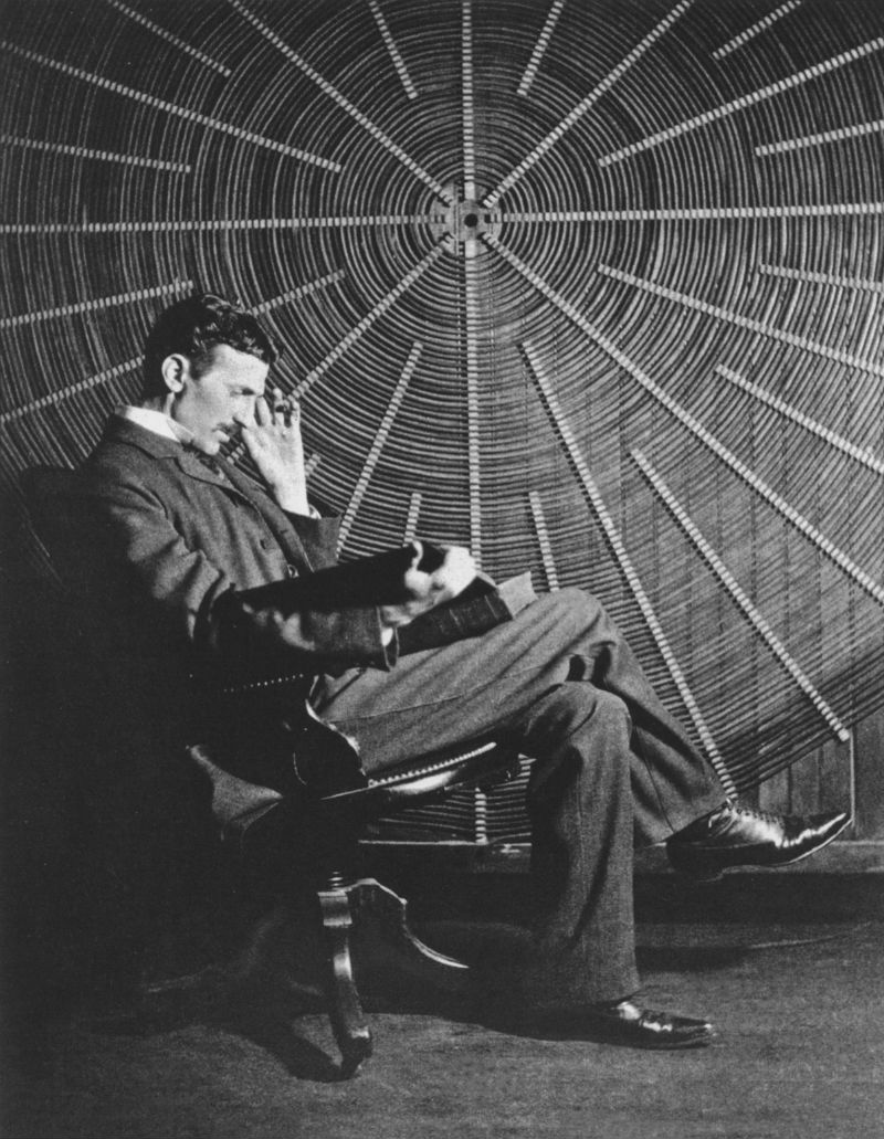
The man himself, Nikola Tesla.

Nuclear magnetic resonance (NMR) was first observed in by Columbia University professor Isidor Rabi in 1937. He was able to detect and measure single states of rotation of atoms and molecules, which measured the magnetic resonance in liquids and solids. Within a decade, scientists were using NMR as standard substance analysis. The first one-dimensional MR image was then made in 1950 by American physicist Herman Carr.

In 1971, Brooklyn’s Downstate Medical Center physician Raymond Damadian discovered that the hydrogen signal in cancerous tissue differs from healthy tissue, because tumors contain more water (i.e., more hydrogen atoms.) This was a huge breakthrough – when an NMR machine was turned off, the radio waves from cancerous tissue remain longer than in healthy tissue. Damadian applied for a patent to use NMR for this purpose in 1972, detailing a hypothetical magnetic resonance machine. A year later, the first NMR image (of a test tube) was made by State University of New York chemist Paul Lauterbur.

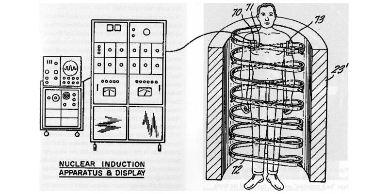
Raymond Damadian’s “Apparatus and method for detecting cancer in tissue.”

Damadian went on to build the first MRI scanner – the “Indomitable” – in 1977, assisted by his post-doctoral students, Michael Goldsmith and Larry Minkoff. The first MRI scan of a healthy human body was made that same year, and the first of a human body with cancer was made a year later.

During that same year (1978) physicist and University of Nottingham professor Peter Mansfield invented the echo-planar imaging (EPI) technique, which cut MR scans down from hours to seconds, and produced clearer images. 

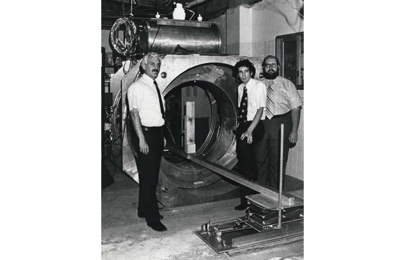
Raymond Damadian, Larry Minkoff and Michael Goldsmith with “Indomitable” (Credit: FONAR)

1978 seems to be a prolific year for the MRI machine! In that year Damadian founded the FONAR corporation, which manufactured the first commercial MRI scanner in 1980. The FONAR corporation scanner was approved by the FDA in 1984.

Real time MR imaging of the heart was developed in 1987. It was initially quite difficult to image the heart because of respiratory and cardiac motion, but cardiac electrocardiograph (ECG) gating, faster scan techniques, and “breath hold” imaging made it possible. Following the development of imaging of axonal transport of supermagnetic metal oxide particles to image neural tracts, functional MRI (fMRI) of the brain was introduced in 1993.

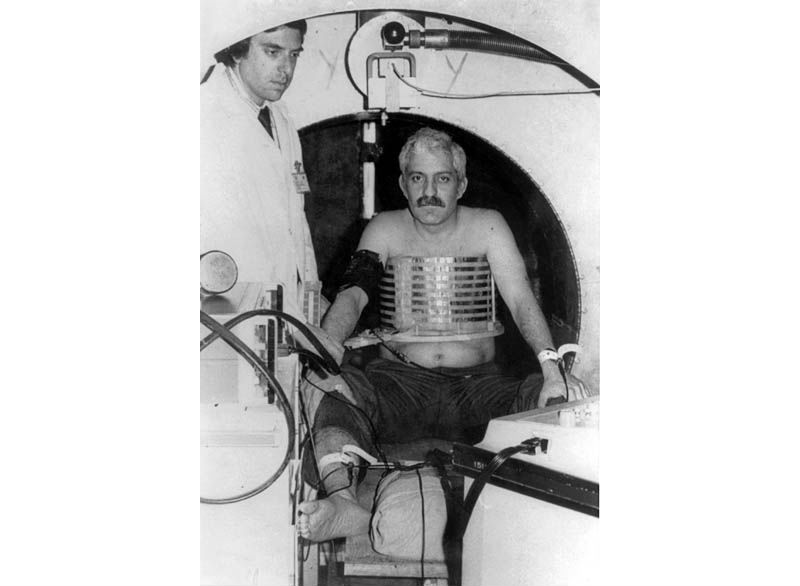
The first (unsuccessful) scan of Damadian. Apparently he was too fat for the NMR receiver coil. (Credit: FONAR)

Since then, great strides have been made in Cardiac and Body MRI, fetal imaging, and fMRI. They are now routine practices in many imaging centers. MRI is usually performed at 1.5T, but higher fields like 3T are gaining popularity because their sensitivity and resolution are increased. Most recently, a combination MRI/radiotherapy machine is currently being built in the University Medical Center in Utrecht – a clinical prototype is expected next year.

Oh, and a friendly reminder. Should you have to undergo an MRI scan, make sure all magnetic materials are safely stowed far away from the machine. Otherwise something like this might happen…

or this…

…or this. 

Related Articles Read More >

An illustration of Embolization Inc.'s Nitinol Enhanced Device (NED).
This nitinol vascular embolization device has another shape memory material up its sleeve
A photo of nitinol, a nickel-titanium alloy used for medical devices such as stents, heart valves, catheters and orthopedics.
What is nitinol and where is it used?
July 2025 edition: The Surgical Robotics issue, featuring Capstan Medical, J&J and Zimmer Biomet
A photo of Capstan Medical's mitral valve implant, which uses nitinol.
Capstan Medical’s R&D head discusses the heart valve and robotics startup’s tech, engineering challenges and solutions, advice for others in medtech and how to join his team
“mdo
EXPAND YOUR KNOWLEDGE AND STAY CONNECTED
Get the latest medical device business news, application and technology trends.

DeviceTalks Weekly

See More >

MDO Digital Edition

Digital Edition

Subscribe to Medical Design & Outsourcing. Bookmark, share and interact with the leading medical design engineering magazine today.

MEDTECH 100 INDEX

Medtech 100 logo
Market Summary > Current Price
The MedTech 100 is a financial index calculated using the BIG100 companies covered in Medical Design and Outsourcing.
DeviceTalks

DeviceTalks is a conversation among medical technology leaders. It's events, podcasts, webinars and one-on-one exchanges of ideas & insights.

DeviceTalks

New MedTech Resource

Medical Tubing

MassDevice

Mass Device

The Medical Device Business Journal. MassDevice is the leading medical device news business journal telling the stories of the devices that save lives.

Visit Website
MDO ad
Medical Design and Outsourcing
  • MassDevice
  • DeviceTalks
  • MedTech100 Index
  • Medical Tubing + Extrusion
  • Medical Design Sourcing
  • Drug Delivery Business News
  • Drug Discovery & Development
  • Pharmaceutical Processing World
  • R&D World
  • About Us/Contact
  • Advertise With Us
  • Subscribe to Print Magazine
  • Subscribe to our E-Newsletter
  • Listen to our Weekly Podcasts
  • Join our DeviceTalks Tuesdays Discussion

Copyright © 2025 WTWH Media, LLC. All Rights Reserved. The material on this site may not be reproduced, distributed, transmitted, cached or otherwise used, except with the prior written permission of WTWH Media LLC. Site Map | Privacy Policy | RSS

Search Medical Design & Outsourcing

  • Home
  • Medical Device Business
    • Mergers & Acquisitions
    • Financial
    • Regulatory
  • Applications
    • Cardiovascular
    • Devices
    • Imaging
    • Implantables
    • Medical Equipment
    • Orthopedic
    • Surgical
  • Technologies
    • Supplies and Components Index
    • Contract Manufacturing
    • Components
    • Electronics
    • Extrusions
    • Materials
    • Motion Control
    • Prototyping
    • Pumps
    • Tubing
  • MedTech Resources
    • Medtech Events in 2025
    • The 2024 Medtech Big 100
    • Medical Device Handbook
    • MedTech 100 Index
    • Subscribe to Print Magazine
    • DeviceTalks
    • Digital Editions
    • eBooks
    • Educational Assets
    • Manufacturer Search
    • Podcasts
    • Print Subscription
    • Webinars / Digital Events
    • Whitepapers
    • Voices
    • Views
    • Video
  • 2025 Leadership
    • 2024 Winners
    • 2023 Winners
    • 2022 Winners
    • 2021 Winners
  • Women in Medtech
  • Advertise
  • Subscribe