Neuroscientists at Cold Spring Harbor Laboratory (CSHL)
reached an important milestone today, publicly releasing the first installment
out of 500 terabytes of data so far collected in their pathbreaking project to
construct the first whole-brain wiring diagram of a vertebrate brain, that of
the mouse.
The data consist of gigapixel images (each close to 1
billion pixels) of whole-brain sections that can be zoomed to show individual
neurons and their processes, providing a “virtual microscope.” The
images are integrated with other data sources from the web, and are being made
fully accessible to neuroscientists as well as interested members of the
general public (http://mouse.brainarchitecture.org).
The data are being released pre-publication in the spirit of open science
initiatives that have become familiar in digital astronomy (e.g., Sloan Digital
Sky Survey) but are not yet as widespread in neurobiology.
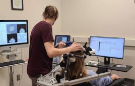
Each sampled brain is represented in about 500 images, each
image showing an optical section through a 20 micron-thick slice of brain
tissue. A multi-resolution viewer permits users to journey through each brain
from “front” to “back,” and thus enables them to follow the
pathways taken through three-dimensional brain space by tracer-labeled neuronal
pathways. The tracers were picked to follow neuronal inputs and outputs of
given brain regions.
“We’re executing a grid-based “shotgun”
strategy for neuronal tract tracing that we first proposed a few years ago, and
which I am pleased to note has gained acceptance elsewhere within the
neuroscience community,” says Partha P. Mitra, Ph.D., the Crick-Clay
Professor of Biomathematics at CSHL and director of the Mouse Brain
Architecture (MBA) Project. After the initial June 1 release, project data will
be made public continuously on a monthly basis, Mitra says.
Project addresses a large gap in knowledge
“Our project seeks to address a remarkable gap in our knowledge of the
brain,” Mitra explains. “Our knowledge of how the brain is wired
remains piecemeal and partial after a century of intense activity. Francis
Crick and Ted Jones emphasized this in an article published in Nature nearly 20
years ago. Yet to understand how the brain works (or fails to work in
neurological or neuropsychiatric disease), it is critical that we understand
this wiring diagram more fully. Further, there remain fundamental questions
about brain evolution that cannot be addressed without obtaining such wiring
diagrams for the brains of different species.”
The MBA Project, which has received critical funding from
the Keck Foundation and from the National Institutes of Health, is
distinguished by the approach advocated by Mitra and colleagues in a position
paper published in 2009. Mitra there proposed mapping vertebrate brains at what
he calls the “mesoscopic” scale, a middle-range amenable to light
microscopy, providing far more detail than, for instance, MRI-based methods,
and yet considerably less detail than is achievable via electron microscopy
(EM). The latter approach, while useful for mapping synaptic connections
between individual neurons, is feasible on a whole-brain basis only for very
small brains (e.g. that of the fruitfly) or very small portions of the mouse
brain.
The pragmatic approach Mitra advocated and which is realized
in this first data release, is to image whole mouse brains in a semi-automated,
quality-controlled process using light microscopy and injected neural tracers (both
viruses and classically used tracer substances). While the basic methodology
has been available for some time, systematically applying it to a grid of
locations spanning the entire brain, and digitizing and re-assembling the
resulting collection of brains, is a new approach made feasible by the rapidly
falling costs of computer storage. A single mouse brain at light-microscope
resolution produces about a terabyte (1 trillion bytes, or 1000 GB) of data;
thus, generating and storing the data set currently being gathered would have
been prohibitively expensive a decade or so ago.
Assembling the circuit diagram at a mesoscopic scale using
‘shotgun approach’
A key point is that at the mesoscopic scale, the team expects to assemble a
picture of connections that are stereotypical – that is, essentially the same
in different individuals, and probably genetically determined in a
species-specific manner. By dividing the volume of a hemisphere of the mouse
brain into 250 equidistant, predefined grid-points, and administering four
different kinds of tracer injections at each grid point — in different animals
of the same sex and age — the eight-member team at CSHL assisted by
collaborating scientists at Boston University, MIT and the University of
California, San Diego seeks to assemble a complete wiring diagram that will be
stitched together from the full dataset.
The project in this sense is analogous to the Human Genome
Project’s “shotgun” approach, in that its final product – a
comprehensive wiring diagram – will be the product of many individually
obtained data components, woven together thanks to the power of advanced
computing and informatics. Indeed, Mitra says one of the genome project’s early
advocates, Dr. James D. Watson (now CSHL chairman emeritus), provided him with
motivation and encouragement to pursue the project.
“We will never understand how the brain works until we
have the wiring diagram,” Dr. Watson comments today. “Mitra is on the
right track and I’m impressed he’s gone from conception to putting out data in
a couple of years on a quite modest budget. His approach deserves strong
funding support.”
The MBA Project was also inspired by early efforts of the
Allen Institute, funded by Microsoft co-founder and philanthropist Paul Allen,
which resulted in assembly of a comprehensive map of gene expression across the
mouse brain. That effort was the product of standard molecular biology
procedures iterated in a quasi-industrialized process. The resulting
whole-brain gene-expression map, while a triumph, was not designed to shed
light on connections in the brain, which became a point of departure for Mitra.
Since the 2009 publication of Mitra and colleagues’ proposal
for meso-scale circuit-mapping projects for whole vertebrate brains, the
approach has not only spawned Mitra’s CSHL project, but also other meso-scale
circuit-mapping projects for the mouse at the Allen Institute and at UCLA. Each
differs in aim and technical detail.
A number of features distinguish the “meso-scale”
circuit project at CSHL. The 20-micron spacing between brain “slices”
gives the CSHL results a particularly rich sense of three-dimensional depth and
detail. The team’s use of four tracers including both classical tracer
substances as well as neurotropic viruses (attenuated or disabled viruses that
infect nerve cells), provides redundancy and helps control for differing
efficacies of the different tracer substances. The images one sees on the MBA
Project website begininng today provide hard data on actual neuronal processes
– the “ground truth” of neuroanatomy, in Mitra’s words — and do not
rely on inferential methodologies such as functional MRI scans and diffusion
tensor imaging to suggest areas in which connections occur. Finally, it is
noteworthy that the slides generated by the project are being physically
stored, to permit re-examination at a later date, using more refined imaging
methods if necessary or as new methods become available.
“Our project is what I’d call a necessary first step in
a much larger enterprise, that of understanding both structure and dynamics of
the vertebrate, and ultimately, the human brain,” says Mitra. “While
facile comparisons with Genome projects should be avoided, the data sets
generated by the MBA and similar projects will provide a useful framework – not
unlike a reference genome – on which we can ‘hang’ all kinds of neuroscience
knowledge, the body of which has always been notably fragmentary.”