Medical Design and Outsourcing

  • Home
  • Medical Device Business
    • Mergers & Acquisitions
    • Financial
    • Regulatory
  • Applications
    • Cardiovascular
    • Devices
    • Imaging
    • Implantables
    • Medical Equipment
    • Orthopedic
    • Surgical
  • Technologies
    • Supplies and Components Index
    • Contract Manufacturing
    • Components
    • Electronics
    • Extrusions
    • Materials
    • Motion Control
    • Prototyping
    • Pumps
    • Tubing
  • MedTech Resources
    • Medtech Events in 2026
    • The 2026 Medtech Big 100
    • Medical Device Handbook
    • MedTech 100 Index
    • Subscribe to Print Magazine
    • DeviceTalks
    • Digital Editions
    • eBooks
    • Educational Assets
    • Manufacturer Search
    • Podcasts
    • Webinars / Digital Events
    • Whitepapers
    • Voices
    • Views
    • Video
  • 2026 Leadership
    • 2025 Winners
    • 2024 Winners
    • 2023 Winners
    • 2022 Winners
    • 2021 Winners
  • Women in Medtech
  • Advertise
  • Subscribe

New Imaging Technique Peers Inside Living Cells

November 17, 2017 By Northwestern University

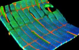
(Credit: Wikimedia Commons)

To undergo high-resolution imaging, cells often must be sliced and diced, dehydrated, painted with toxic stains, or embedded in resin. For cells, the result is certain death.

But if researchers can only view the inner workings of dead cells, they’re only seeing part of the story. They cannot monitor living cells’ dynamic real-time processes, such as metabolic reactions or responses to diseases or treatments.

“Sub-cellular components and structures have a profound influence on the behavior of the complex cellular machinery and systems biology,” said Northwestern University’s Gajendra Shekhawat. “However, unraveling the structures and components inside the cell is very challenging because they are so fragile.”

Now Shekhawat and Vinayak P. Dravid, the Abraham Harris Professor of Materials Science and Engineering at Northwestern Engineering, have developed a novel non-invasive imaging system that makes it possible to view the sub-cellular architecture of live cells at nanometer-scale resolution. Called Ultrasound Bioprobe, the technique combines ultrasound waves with atomic force microscopy, interacting with live cells to determine the changes in their mechanical behavior.

Supported by the National Science Foundation (NSF) and the National Heart, Lung, and Blood Institute, the research was recently published in Science Advances. Shekhawat and Dravid served as the paper’s co-corresponding authors. Shekhawat, a research associate professor in materials science and engineering, was also the first author of the paper. The research was completed in the Northwestern University Atomic and Nanoscale Characterization Experimental (NUANCE) Center. NUANCE is the lead facility in the NSF-supported National Nanotechnology Coordinated Infrastructure (NNCI) Program, which is headquartered at Northwestern and called the Soft and Hybrid Nanotechnology Experimental (SHyNE) Resource.

Despite recent advances in imaging, there is currently no single method that provides high-resolution and high-sensitivity images of living sub-cellular structures. Fluorescent and confocal microscopy, which are traditional methods for monitoring the biological interactions inside cells, suffer from poor spatial resolution and require invasive dyes or labels to enhance contrast and highlight structures within biological tissues. Light and acoustic wave imaging are unable to view structures smaller than a few hundred nanometers. Scanning probe microscopy can provide very high spatial resolution but can only identify surface structures rather than peer inside a cell. And while electron microscopy can view fine details at the sub-cellular level, it’s a destructive technique that cannot be used for living biological tissues.

“Many roadblocks have existed,” said Dravid, who directs the NUANCE Center and the SHyNE Resource. “Characterization of the complex dynamics of biological processes, especially signal pathways at nanoscale resolution, has remained a challenge.”

Shekhawat and Dravid’s Ultrasound Bioprobe, however, bypasses these issues. Its ultrasound waves non-invasively image deeply buried intracellular features. And its atomic force microscopy probe provides high sensitivity and mechanical contrast of the scattered ultrasound waves. The result? Non-destructive, remarkably high-contrast, nanoscale images of structures and components deep inside living tissues and cells.

“Using this non-invasive approach, we can monitor real-time imaging of the nanomechanical changes in complex biological systems,” Shekhawat said. “This could provide clues for early diagnostics and potential pathways for developing therapeutic strategies.”

Next, the team plans to expand its technique to diverse biomedical applications, such as the nanomechanics of soft tissues such as skin, enamels, and bones to probe their three-dimensional architecture down to nanoscale spatial resolution.

“A significant variation in cellular nanostructures and mechanics can be directly influenced by the cancer conditions of a cell,” Dravid said. “So Ultrasound Bioprobe could also expand our fundamental understanding of the nanomechanics at play within cancer cells.”
Related Articles Read More >

The most innovative medical devices of 2026
An illustration showing Corcym's Memo 4D Curve's laser-cut nitinol core.
Laser-cut nitinol and a layered design let these cardiac implants flex
Reconstructed 3D fluorescence image of the stained biohybrid mesh-tissue integration. The red, green, and blue colors correspond to the mesh, F-actin, and nuclei, respectively.
Researchers mesh electronics with human cells to develop living battery
A photo of the Paradromics Connexus brain-computer interface on a fingertip.
How Paradromics designed its Connexus brain-computer interface system
“mdo
EXPAND YOUR KNOWLEDGE AND STAY CONNECTED
Get the latest medical device business news, application and technology trends.

DeviceTalks Weekly

See More >

MDO Digital Edition

Digital Edition

Subscribe to Medical Design & Outsourcing. Bookmark, share and interact with the leading medical design engineering magazine today.

MEDTECH 100 INDEX

Medtech 100 logo
Market Summary > Current Price
The MedTech 100 is a financial index calculated using the BIG100 companies covered in Medical Design and Outsourcing.

New MedTech Resource

Medical Tubing

MassDevice

Mass Device

The Medical Device Business Journal. MassDevice is the leading medical device news business journal telling the stories of the devices that save lives.

Visit Website
MDO ad
Medical Design and Outsourcing
  • MassDevice
  • DeviceTalks
  • MedTech100 Index
  • Medical Tubing + Extrusion
  • Medical Design Sourcing
  • Drug Delivery Business News
  • Drug Discovery & Development
  • Pharmaceutical Processing World
  • R&D World
  • About Us/Contact
  • Advertise With Us
  • Subscribe to Print Magazine
  • Subscribe to our E-Newsletter
  • Listen to our Weekly Podcasts
  • Join our DeviceTalks Tuesdays Discussion

Copyright © 2026 Arrowfly, LLC. All Rights Reserved. The material on this site may not be reproduced, distributed, transmitted, cached or otherwise used, except with the prior written permission of Arrowfly LLC. Site Map | Privacy Policy | RSS

Search Medical Design & Outsourcing

  • Home
  • Medical Device Business
    • Mergers & Acquisitions
    • Financial
    • Regulatory
  • Applications
    • Cardiovascular
    • Devices
    • Imaging
    • Implantables
    • Medical Equipment
    • Orthopedic
    • Surgical
  • Technologies
    • Supplies and Components Index
    • Contract Manufacturing
    • Components
    • Electronics
    • Extrusions
    • Materials
    • Motion Control
    • Prototyping
    • Pumps
    • Tubing
  • MedTech Resources
    • Medtech Events in 2026
    • The 2026 Medtech Big 100
    • Medical Device Handbook
    • MedTech 100 Index
    • Subscribe to Print Magazine
    • DeviceTalks
    • Digital Editions
    • eBooks
    • Educational Assets
    • Manufacturer Search
    • Podcasts
    • Webinars / Digital Events
    • Whitepapers
    • Voices
    • Views
    • Video
  • 2026 Leadership
    • 2025 Winners
    • 2024 Winners
    • 2023 Winners
    • 2022 Winners
    • 2021 Winners
  • Women in Medtech
  • Advertise
  • Subscribe