The early stages of esophageal cancer is difficult to recognize because it can easily be missed. Eindhoven University of Technology in The Netherlands has been working with the Catharina Hospital in Eindhoven to develop a method to enable a computer to scan esophagus images for signs of esophageal cancer.
The program recognizes early cancer with almost as much accuracy as top specialists, according to researchers. The results are published in the article, “Computer-aided detection of early neoplastic lesions in Barrett’s esophagus,” which appears in the July issue of the scientific journal Endoscopy.
People with prolonged reflux tend to develop abnormal tissue in the esophagus due to gastric irritation. This so-called Barrett’s esophagus is one of the major risk factors in the development of esophageal cancer in the Western world and, therefore, people with a Barrett’s esophagus regularly undergo an endoscopic hospital check.
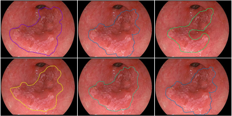
Six contours were drawn by five experts and the computer systems, of early esophageal cancer. The image top right is the contour drawn by the computer system. (Credit: Eindhoven University of Technology)
The earliest stages of esophageal cancer are, however, very difficult to detect. Thus, when the cancer begins, and when it is easy to treat, it can be missed. Once the cancer reaches an advanced stage, the five-year survival rate is less than fifty percent. Reflux often occurs in overweight people, and the incidence of obesity is increasing.
Four years ago Dr. Erik Schoon, gastroenterologist at the Catharina Hospital and top specialist in the area of the Barrett’s esophagus, visited with the Eindhoven Video Coding and Architectures Research Group, which worked together with Schoon to develop new methods that can accurately investigate the first signs of cancer.
The computer analysis should become available in hospitals over time to help gastroenterologists recognize the earliest stages of cancer and so pave the way for 100 percent recognition and treatment, according to the researchers.
That would save many patients from having to undergo surgery that removes part of the esophagus, which normally occurs when the cancer is detected late. The treatment of early cancer is much less invasive for patients and tends to comprise a microsurgical operation from within. It is also much cheaper. Additionally, doctors who are not Barrett specialists can use the method to learn to recognize abnormalities more quickly.
Before the new method can be employed, the software has to be improved and made suitable for analyzing real-time video frames. Then a number of extensive hospital tests will need to follow. It will probably take five to 10 years before broad implementation occurs. Currently, there are various subsidy applications being submitted to fund the project further.
The study is being undertaken by Eindhoven University of Technology in collaboration with the Catharina Hospital Eindhoven, the Leuven University Hospital, the Barmherzige Brüder Regensburg Hospital, the St. Antonius Hospital in Nieuwegein and the Academic Medical Center Amsterdam.


