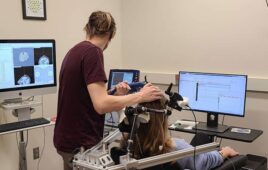
4D flow CMR can be employed to measure in-vivo 3D blood flow dynamics in the heart and atria. (Credit: Northwestern University)
An advancement in stroke prevention has been made. A 4-D brain imaging technique to predict strokes has been developed by Northwestern University professor, Michael Markl.
According to Northwestern, the new MRI technique is designed to measure how quickly blood flows through the vessels. Strokes, which are triggered by blood clots from the heart to the brain, can be better detected with the new 4-D imaging. With the ability to display the images in three special dimensions, the new 4-D technique, also known as “atrial 4-D flow CMR,” is able to show a better image of a person’s blood flow from the heart and the rest of the body. Those who experience atrial fibrillation have a higher chance of experiencing strokes, but now have the opportunity to detect them early.
Specifically, the 4-D MRI program is a software that can be used with existing MRI equipment, therefore hospitals have no need to replace their current systems. No special equipment or equipment upgrades are needed for the software to be used.
Markl stated to Northwestern, “We simply programmed the scanner to generate information differently — in a way that wasn’t previously available. It allows you to measure flow, diffusion of molecules, and tissue elasticity. You can interrogate the human body in a very detailed manner.”
The software was created in the hopes of earlier detection and more precise treatment.