(Credit: TU Delft)
An extra detector on an electron microscope can help determine which molecules are found in which parts of a cell. This is what scientists at the University Medical Center Groningen (UMCG) and Delft University of Technology report in an article published today in the journal Scientific Reports. “This detector enables us to assign a colour to molecules in a cell,” says Ben Giepmans, the team leader from Groningen. “Multicolour electron microscopes are a new addition to medical research, and they could generate interesting results.”
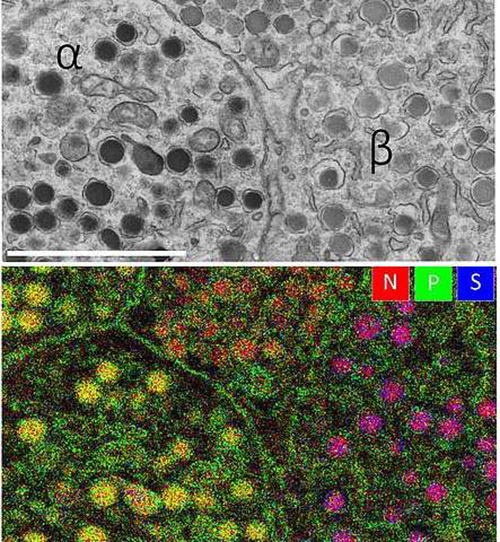
Electron microscopes can zoom in great detail, thus making the tiniest structures in a cell visible. They are therefore much more precise than optical microscopes, which have been around for much longer. “But an electron microscope always shows images in gray scales,” Giepmans explains. “We have now demonstrated that you can introduce colour with this detector. You can compare it with Google Earth—satellite images give a good impression of what a small part of the Earth looks like, but if you colour the roads and cities, it is much easier to find your bearings. Similarly, if you colour molecules, you make it easier to see which biological structures you are looking at.”
Identifying elements
The researchers used a detector that was developed for materials science. The Delft team leader Jacob Hoogenboom says, “We purchased the detector to study extremely small structures for the semiconductor industry. We were already working with the UMCG on other projects. They had used comparable techniques to colour in biological samples, but this only produced two colours. So we thought we’d study them with this detector, too.” The detector can identify each separate building block of molecules, including nitrogen, phosphorus, sulphur, iron and other metals. Giepmans says, “DNA contains a lot of phosphorus, for instance. If we map the phosphorus in a cell and assign it a colour, we can see where the DNA is.”
Application
The researchers applied the technique to their own field of research, type 1 diabetes. “
We looked at the cells in the pancreas of a rat that was sensitive to type 1 diabetes. We could clearly identify the different cells in the pancreas. Insulin-producing cells acquired a colour from the sulphur, because insulin contains a lot of sulphur, whereas cells that produce glucagon took on another colour, because that hormone contains other elements.”
Tissue was identified in Groningen and sent to Delft, where the new detector was used to analyse certain regions. This led to surprising observations. “In this rat, we could see substances in parts of the pancreas where they are not usually found,” Giepmans explains. The UMCG now has its own ‘colour EM’ detector, and Giepmans is already receiving cell material from home and abroad so that he can test the new technique.
The researchers are not the first to colour elements using an electron microscope. “In a previous study, they could only colour two substances. We can now measure and colour many different elements at the same time. I knew that it must be possible. I dreamt about it for a long time, but it only got off the ground when we started working with Delft and used their detector on our tissue.” Interdisciplinary collaboration led to concrete results.
“What is perhaps best about this technique is that it is affordable. It really is a new microscopy tool that we are already using for many research groups.”


