Using high-resolution electron microscopy, Columbia University Medical Center (CUMC) researchers have uncovered new details of the structure and function of an intracellular channel that controls the contraction of skeletal muscle. The findings, published in Cell, could lead to new treatments for a variety of muscle disorders.
“It’s exciting to see this channel in such detail. From this information, we can learn more about how the channel functions in normal conditions and how it malfunctions in disease states. Most important, these insights greatly improve our ability to design new drugs to treat a range of muscle diseases,” said co-study leader Andrew R. Marks, M.D., the Clyde ’56 and Helen Wu Professor of Molecular Cardiology (in Medicine), professor and chair of physiology and cellular biophysics, and founding director of the Clyde and Helen Wu Center for Molecular Cardiology at CUMC.
Ryanodine receptors (RyRs)/calcium release channels—first visualized in 1988 by Joachim Frank, Ph.D., using electron microscopy and cloned in 1989 by Dr. Marks—belong to a family of intracellular channels that control muscle contraction. The two major types, RyR1s and RyR2s, are found in skeletal muscle cells and heart muscle cells, respectively. Leaky RyRs have been implicated in a variety of conditions, including age-related muscle weakness, muscular dystrophy, and heart failure.
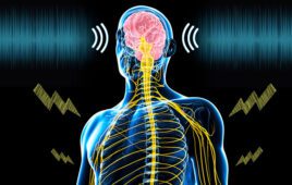
Movement begins when the brain sends a signal to a skeletal muscle cell, changing the voltage across the cell membrane. The voltage change activates RyR1 channels, which are located in a calcium-storage organelle called the sarco/endoplasmic reticulum. When activated, the channels open the organelle’s floodgates and release calcium into the cell body, triggering muscle contraction.
In a previous study, published in Nature, Dr. Marks and his team used an advanced electron microscopy (EM) technique called cryo-EM, combined with computer modeling, to determine the 3D structure of the RyR1 channel in its closed state (in which no calcium is allowed out of the sarco/endoplasmic reticulum).
The current study provides the first images of the RyR1 channel in the open state, as well as in various conformations in which it has changed shape but the channel has not yet opened. “Now, rather than having a static picture of the channel, we can make movies of a sort showing how various parts of the molecule are moving around,” said co-study leader Dr. Frank, professor of biochemistry and molecular biophysics and of biological sciences at CUMC, a Howard Hughes Medical Institute (HHMI) investigator, and a world-leading expert in electron microscopy.
Co-first author Oliver B. Clarke, Ph.D., a postdoctoral fellow at CUMC at the time of the study and now an associate research scientist in the Department of Biochemistry and Molecular Biophysics at CUMC, said, “with this information, we can begin to understand how ligands regulate ryanodine receptor activity. This may eventually help us develop improved therapeutics to treat diseases where RyR-mediated Ca2+ ‘leak’ is a contributing factor.”
The researchers were also able to visualize where and how calcium, adenosine triphosphate, and caffeine—the primary molecules that activate RyR1—bind to the channels.
Dr. Marks has already developed drugs that target malfunctioning RyR channels. Studies have shown that these drugs can increase muscle force in animals with age-related muscle weakness or Duchenne muscular dystrophy and improve heart function in animals with heart failure. Clinical trials to test the drugs in children with Duchenne muscular dystrophy and RyR1 myopathy may begin as early as next year.
The other co-study leader is Wayne A. Hendrickson, Ph.D., university professor in the Department of Biochemistry and Molecular Biophysics and Violin Family Professor of Physiology and Cellular Biophysics at CUMC and an eminent X-ray crystallographer.
The other co-first authors are Amédée des Georges, Ph.D., and Ran Zalk, Ph.D., both postdoctoral fellows at CUMC at the time of the study. Dr. des Georges is now at CUNY Advanced Science Research Center, New York, NY, and Dr. Zalk is at the National Institute for Biotechnology in the Negev, Ben-Gurion University of the Negev, Beer Sheva, Israel.
The paper is titled, “Structural basis for gating and activation of RyR1.” The other contributors are: Qi Yuan, Kendall J. Condon, and Robert A. Grassucci (all at CUMC).
This study was supported by the National Institutes of Health (R01AR060037, R01HL061503, P41GM11679, R01GM29169), HHMI, the American Heart Association, and the Charles H. Revson Foundation.
Dr. Marks is a consultant and board member of ARMGO Pharma, Inc., which is targeting RyR channels for therapeutic purposes. The other authors declare no financial or other conflicts of interest.
Columbia University Medical Center provides international leadership in basic, preclinical, and clinical research; medical and health sciences education; and patient care. The medical center trains future leaders and includes the dedicated work of many physicians, scientists, public health professionals, dentists, and nurses at the College of Physicians and Surgeons, the Mailman School of Public Health, the College of Dental Medicine, the School of Nursing, the biomedical departments of the Graduate School of Arts and Sciences, and allied research centers and institutions. Columbia University Medical Center is home to the largest medical research enterprise in New York City and State and one of the largest faculty medical practices in the Northeast. For more information: cumc.columbia.edu or columbiadoctors.org.
(Source: Newswise)


