South Jersey Radiology Associates (SJRA) is transforming prostate cancer screening by putting non-invasive 3T magnetic resonance imaging technology into the hands of imaging specialists.
Because the 3T magnet is so powerful, the procedure does not require the invasive endo-rectal coil that 1.5T MRI machines require. Patient comfort – and willingness to undergo the imaging procedure – is dramatically improved. And there is no radiation exposure with MR imaging.
For all these reasons, Dr. Gregory J. Goodworth, MD, DABR, a body imaging specialist at SJRA, predicts the non-invasive prostate scan will be as widely known as the mammogram or colonoscopy in the very near future.
“The first prostate MRIs were performed more than 15 years ago using 1.5T machines (T, for Tesla, refers to the unit of measurement quantifying the strength of a magnetic field). Besides requiring the invasive, endo-rectal coil, the 1.5T machines produced images that were “not very specific or sensitive,” Goodworth said. “If there was a big tumor, you might see it, but rarely would you see small disease.”
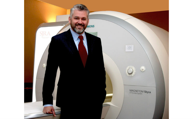
Dr. Gregory J. Goodworth with the 3T MRI Skyra (Credit: South Jersey Radiology Associates)
By comparison, the 3T MRI’s powerful magnet allows imaging of very small body parts at very high resolution, down to a few millimeters.
He compared the evolution of 3T MRI to cell phone cameras. “Your first cell phone had a low-megapixel camera that produced low quality, grainy images,” Goodworth said. “Compare that to cell phone cameras today and the crisp, high-resolution images that they produce.”
Before MR imaging of the prostate became available, urologists had just three tools available to evaluate prostate disease in men: The digital rectal exam, the PSA test (a blood test that measures the level of prostate-specific antigen), and the transrectal ultrasound-guided biopsy.
In the latter procedure, needles are inserted through a rectal probe into the prostate to sample areas of the gland. Goodworth noted that this highly invasive procedure is also highly inaccurate because about 80 percent of the prostate gland goes completely unevaluated. With the 3T MRI, “I can see the whole prostate gland, including those areas that transrectal biopsy needles can’t reach,” he said.
For patients who have had prior negative biopsy results but are at risk for developing cancer, 3T MRI allows monitoring of prostate changes without subjecting the patient to repeated biopsies.
And for patients with high PSA levels or a non-life threatening prostate tumor, 3T MRI is a very powerful tool for avoiding overtreatment and the related potential risks from surgery or radiation, such as incontinence, pain, and erectile dysfunction, Goodworth added. This is particularly important, since most men diagnosed with prostate cancer will not die from it.


