Aptyx Senior Engineer Daniel Meckes [Photo courtesy of Aptyx]
When I first began researching pulsed-field ablation (PFA) about seven years ago, I encountered a surprising amount of oversimplified information in the literature.
PFA was often described as using low-voltage pulses (around 500 V), being entirely nonthermal, not requiring electrode–tissue contact, and exhibiting cardiac tissue selectivity. The explanation I heard at the time made it sound almost effortless: an ablation catheter you could simply move around the heart to ablate exactly the tissue you wanted, with minimal risk to the patient.
It sounded compelling, but likely too good to be true. So, I set out to investigate.
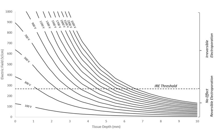
Over the next couple of years, I found that PFA actually requires much higher voltages than commonly reported at the time, depends heavily on good electrode–tissue contact, can become dangerously thermal without proper waveform design, and relies on a far less rigorous notion of tissue selectivity tied to irreversible electroporation (IRE) thresholds.
Register now: The MDO Min-Vasive Medtech webinar series returns Aug. 18-20
PFA development challenges
The first problem I tackled in developing a PFA system was lesion depth. Using computational models based on Maxwell’s equations, I showed that electric field volume scales with applied voltage: lower voltage produces smaller field volumes, while higher voltage expands them. This indicated that achieving clinically relevant lesion depths throughout the heart would require far more than 500 V.
[Illustration courtesy of Dan Meckes/Aptyx]
At those voltages however, thermal effects became an obvious concern. I expanded my models to include heat transfer in cardiac tissue, accounting for cooling from blood flow.
The results were alarming. I measured highly localized, instantaneous temperature spikes exceeding 400°C. I initially assumed there must be an error in my model, but after extensive verification, the results held. This led me to systematically tune waveform parameters such as pulse duration, pulse count, and inter-pulse delays until I achieved thermally safe conditions. Still, translating these rapid temperature spikes into a meaningful nonthermal design constraint remained challenging.

[Illustration courtesy of Dan Meckes/Aptyx]
Investigating thermal damage
Further research showed that cardiac tissue begins to experience thermal damage around 50°C after approximately 25 seconds. Using this, I developed a method to normalize the transient temperature spikes in my model to an equivalent constant-temperature exposure, allowing me to align my designs with established thermal damage thresholds. With that, I established a framework for designing a high-voltage yet clinically nonthermal PFA waveforms.
I then tested hundreds of waveform variations in simulation and found that at high voltages, the energy becomes extremely sensitive. Very fast (nanosecond) pulses were required and small changes in waveform parameters can produce dangerously high thermal doses. It became clear that reaching this level of understanding without numerical modeling would be extraordinarily difficult, if not impossible.
Another major challenge was the reported IRE thresholds for cardiac tissue, which are often referenced for tissue selectivity. In practice, these thresholds are not determined purely mathematically, but rely on comparing experimental outcomes with model-predicted electric field distributions. If the model is inaccurate, the derived threshold is as well. Compounding this, many models incorporate an electric field “growth factor” to account for increased tissue conductivity that occurs during PFA.
In the literature, however, I observed models inflating fields by as much as eight- to ten-fold, suggesting that, rather than determining true thresholds, results may be highly dependent on model assumptions. This introduces additional uncertainty into what is often treated as a purely quantitative process. In practice, this can make threshold values difficult to interpret with confidence.
The results
Ultimately, what matters is whether a safe and effective PFA system can be built. During my time, there wasn’t any concern regarding hemolysis, which is a serious complication being reported today. Reflecting on this now, one potential contributor may be instantaneous temperature spikes associated with suboptimal waveform design.
I worked on this problem as lead engineer within a small startup, without the time or resources available to larger companies currently dominating the market. Despite that, I developed a first prototype in a couple of years.
It’s a high-voltage PFA system capable of delivering nanosecond pulses. It is now undergoing clinical trials in Australia. This was accomplished by focusing on the underlying physics and validating assumptions through modeling and iteration.
Numerical modeling was central to that effort, enabling precise control, rapid exploration and insights that would have been difficult to obtain otherwise. The result was a more controlled, predictable approach to PFA design, grounded in early engineering decisions and rigorous validation.
Daniel Meckes is an R&D engineer at Aptyx with 12 years of experience in design and development of advanced catheters and implantable systems. His work balances first principal engineering and physical prototypes to streamline concept ideation and the engineering process.
Read more MDO Contributions and learn how to submit your own.
The opinions expressed in this blog post are the author’s only and do not necessarily reflect those of Medical Design & Outsourcing or its employees.