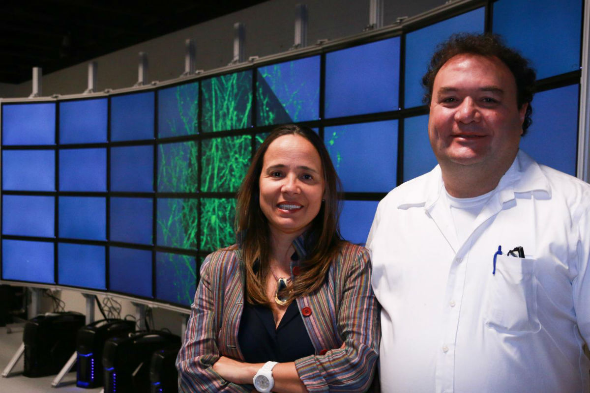
Alessandra Angelucci (left), a University of Utah professor of ophthalmology and visual science, and University of Utah computer science professor Valerio Pascucci stand in front of a computer model of a cluster of neurons scanned from a primate’s brain. They have developed software that maps out a monkey’s brain and more easily creates a 3-D model, providing a more complete picture of how the brain is wired. This method of visualizing the brain could help medical researchers better understand what happens to the human brain after it is affected by conditions such as autism, depression, anxiety or retinal degeneration. (Credit: University of Utah College of Engineering/Dan Hixson)
The animal brain is so complex, it would take a supercomputer and vast amounts of data to create a detailed 3D model of the billions of neurons that power it.
But computer scientists and a professor of ophthalmology at the University of Utah have developed software that maps out a monkey’s brain and more easily creates a 3D model, providing a more complete picture of how the brain is wired. Their process was announced this week at Neuroscience 2015, the annual Society for Neuroscience meeting in Chicago.
“If you understand how things are wired in the normal brain, you can use this as a basis to understand how these connections are disrupted in the abnormal brain,” said Alessandra Angelucci, professor of ophthalmology and visual science at the University of Utah.
Getting a more accurate view of the brain’s network of neurons can help medical researchers understand how the brain’s connectivity is disrupted in mental and neurological conditions such as schizophrenia, depression, anxiety and autism. For Angelucci, who works at the University of Utah’s Moran Eye Center, this also can aid research on such vision-related conditions as amblyopia, a disorder where one or both eyes lack visual acuity, and various forms of retinal degeneration. Angelucci has been using this software on a monkey’s brain because it most closely resembles the human brain.
Alessandra Angelucci (right), a University of Utah professor of ophthalmology and visual science, and University of Utah computer science professor Valerio Pascucci stand in front of a computer model of a cluster of neurons scanned from a primate’s brain. They have developed software that maps out a monkey’s brain and more easily creates a 3-D model, providing a more complete picture of how the brain is wired. This method of visualizing the brain could help medical researchers better understand what happens to the human brain after it is affected by conditions such as autism, depression, anxiety or retinal degeneration. (Credit: University of Utah College of Engineering/Dan Hixson)
In the past, researchers would have to scan thousands of thin layers of a primate’s brain through a microscope in order to get a view of its neurons, the brain’s cells that transmit nerve impulses. There was no practical way to make a 3D model of the brain from these layers. For example, a high-resolution scan of a part of the brain the size of a penny would generate about two million images all totaling 30 terabytes (30,000 gigabytes) in files.
“It takes a lot of computer power because we now have to reconstruct a three-dimensional image out of this — thousands and thousands of images of tissue,” Angelucci said. “It was simply impossible because there is no computer or software that can handle that. It involves terabytes and terabytes of data.”
A team led by Valerio Pascucci, a professor in the University of Utah’s School of Computing and director of the university’s Center for Extreme Data Management Analysis and Visualization (CEDMAV) at the Scientific Computing and Imaging Institute (SCI), has developed software that can create a 3D model of an animal’s brain that is much quicker and requires less computer power and system memory.
The team took an existing software platform CEDMAV created called VISUS (Visualization Streams for Ultimate Scalability) and adapted it to assemble high-resolution images of different sections of the brain into one 3D model that can be viewed at different angles. VISUS is used to visualize huge sets of data to create weather or energy simulations or high-resolution images of cities.
To create images of a brain, researchers first use a new method known as CLARITY that makes the brain tissue transparent by immersing it in special hydrogels. With the new software, hundreds of 3D blocks of the brain are then scanned one at a time with a two-photon microscope, and scientists can view the scans immediately as opposed to waiting for them to download.
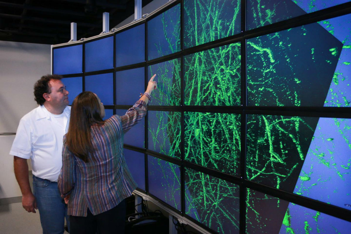
With the help of a researcher, the software then can more easily and quickly assemble the blocks into one complete picture of a region of the brain and create a 3D model that allows the scientist to view areas and angles that couldn’t be seen as easily with 2-D images. In this way, researchers can map out the individual neurons and their long tails, known as axons.
“It really unleashes a different level of understanding of the data itself — being able to look at something fully in 3D and to rotate and look at in front and in back,” Pascucci said. “We have seen over and over in many fields that this makes people understand more quickly and much better the spatial relationship among all the parts.”
With the software, researchers also can monitor the scanning of the brain — which can take weeks — and make sure that no bad images are created in the process, saving precious time.
Thanks to this new tool, medical researchers can now study and better understand how the brain’s connectivity is disrupted in abnormal conditions, for example what happens to the brain’s neural network resulting from retinal degeneration or conditions such as autism.
“We can view it, reconstruct it and understand its connectivity,” Angelucci said. “This software speeds up our ability to do that.”
Also working with Pascucci on the software is master’s students Cameron Christensen and Mike Liu, graduate research assistant Duong Hoang and SCI senior engineer Giorgio Scorzelli. Working with Angelucci is Frederick Federer, a senior postdoctoral fellow. The research was supported by funds from the National Institutes of Health, the National Science Foundation and Research to Prevent Blindness.