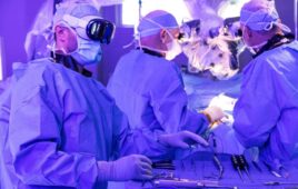
For the first time, surgeons at Johns Hopkins Hospital were able to harness real-time image guidance during a robotic spine surgery procedure to insert screws into a patient’s spine.
A new robot invented by Johns Hopkins neurosurgeon Nicholas Theodore uses real-time imaging during spine surgeries to point to a spot and insert a screw on that same spot.
The process offers a marked improvement over the accuracy of placement compared with other image-guided procedures.
Dr. Nicholas Theodore – professor of neurosurgery at the Johns Hopkins University School of Medicine and director of the Neurosurgical Spine Center of Johns Hopkins Medicine – invented the robot and maintains a financial interest in the technology.
According to the hospital, current image-guided surgical procedures require the surgeon to look back and forth between the patient and an image. The delay and change of focus cause imperfection of screw placement. While often these placements are “good enough,” Theodore said he was excited to offer patients submillimeter accuracy in screw placement.
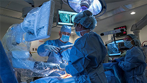
This new robot pairs a CT scan of the patient with the actual patient, allowing the surgeon to point to a spot on the CT scan and tell the robot to aim for that same spot. The robot is also connected to a camera, which reads landmarks on the patient. The robot is able to process what the camera “sees” with the CT image in real time.
The system addresses a big fear in this type of procedure: movement. If the patient breathes or otherwise moves slightly, there can be a change in the placement. The robot at Johns Hopkins can sense changes in position and adjust accordingly.
In the first-case use, a patient suffered a fall at home and opted to use the new robot. According to a video testimonial, that the patient had significant degenerative changes, which would have made routine placement of the screws difficult.