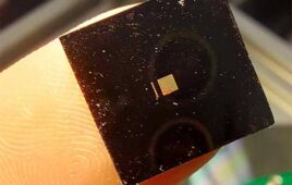
[Image from MIT]
Scientists use implantable fibers to study the brain, giving them the opportunity to stimulate specific parts of the brain to monitor electrical responses. Before the rubber fibers, stiff and brittle fibers could damage spinal cord tissue as the spine flexed.
The new rubber-like fibers can deliver optical impulses for optoelectronic stimulation and electrical connections for stimulation and monitoring.
“I wanted to create a multimodal interface with mechanical properties compatible with tissues, for neural stimulation and recording,” said Alice Lu, MIT graduate student and one of the authors of the study, in a press release.
In order to create the proper tools for understanding spinal cord functions, it was important for the researchers to create a device that could stretch.
“The spinal cord is not only bending but also stretching during movement,” said Lu.
Rubber-like compounds and elastomers are not adaptable to fiber drawing. Since the spine stretches about 12% during normal movements, the need for a rubbery material with the same elasticity was imperative.
“The goal was to mimic the stretchiness and softness and flexibility of the spinal cord, said Polina Anikeeva, one of the authors of the study and a professor at MIT. “You can match the stretchiness with a rubber. But drawing rubber is difficult – most of them just melt.”
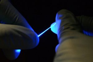
The researchers used a new transparent elastomer as a waveguide for optical signals and a coating of a mesh of silver nanowires to create a conductive layer for the electrical signals. That material was embedded in a polymer coating to process the elastomer and allow it to be drawn into a stretchable and flexible fiber. After the drawing process, the coating dissolves.
What the researchers are left with is a transparent fiber with nanowire coating that is electrically conductive and stretchy. The fiber can stretch 20 to 30% without being affected.
“They’re so floppy, you could use them to do sutures and deliver light at the same time,” said Anikeeva.
“We’re the first to develop something that enables simultaneous electrical recording and optical stimulation in the spinal cords of freely moving mice,” said Lu. “So we hope our work opens up new avenues for neuroscience research.”
Studying the nerves of the spinal cord could lead to better treatments for spinal cord injuries. Scientists currently have to use larger animals in studies because larger nerve fibers are able to experience more rigid wires for stimulation and recording.
“There are many types of cells in the spinal cord, and we don’t know how the different types respond to recovery, or lack of recovery, after an injury,” said Lu.
Spinal cord injuries are rare and affect less than 200,000 of the U.S. population per year, according to the Mayo Clinic. It occurs when any part of the spinal cord or nerves at the end of the spinal canal get damages. The damage often causes someone to experience changes in strength and sensation below the side of the injury.
“Eventually, we’d like to be able to use something like this to combat spinal cord injury. But first, we have to have biocompatibility and to be able to withstand the stresses in the spinal cord without causing any damage,” said Anikeeva.
The research was supported by the National Science Foundation, the National Institute of Neurological Disorders and Stroke, the U.S. Army Research Laboratory and the U.S. Army Research Office through the Institute for Soldier Nanotechnologies at MIT. The researchers published their work in the Science Advances journal.
[Want to stay more on top of MDO content? Subscribe to our weekly e-newsletter.]