A smartphone-based digital ophthalmoscope shows promise as both a teaching tool and an inexpensive, easy-to-use examination instrument for public health services, according to a recent study led by Dr. Claudia Campos at the Wake Forest Baptist Medical Center. The device, known as the D-EYE Smartphone-based Retinal Imaging System, is designed to attach to iOS and Android devices via a specially designed, lightweight bumper. The D-EYE was examined by a team of physicians to determine if the system was a beneficial alternative for residents in comparison to the traditional ophthalmoscope. A poster presentation displaying the results of the study, which concluded that using smartphone technology to perform funduscopic examinations is a promising teaching tool, was given at the Society of General Internal Medicine’s annual conference, held May 11-14 of this year.
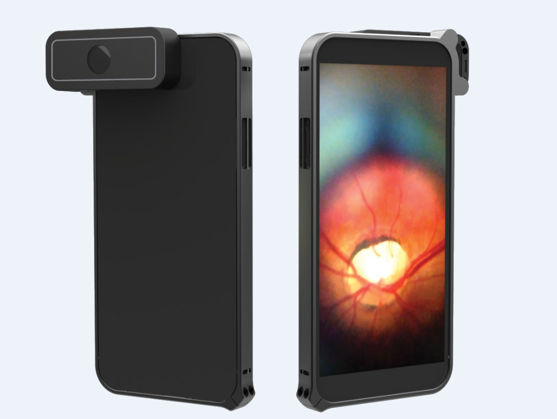
Image courtesy of D-EYE.
D-EYE uses the principle of direct ophthalmology—utilizing the phone’s camera lens and LED light source, which is off-axis from the camera aperture, to illuminate the interior of the eye for examination, resulting in no corneal glare. The examiner focuses the camera lens on the retina and begins the recording and post processing. Examiners will also be able to send the recorded images wirelessly to an optional HIPAA-compliant private and secure cloud-based data and image storage service, for further evaluation and archiving.
When the pupil is dilated, the device captures a field of view of approximately 20 degrees in a single fundus image at a distance of 1 cm from the patient’s eye. When examining an un-dilated eye, the field of view is approximately 5-8 degrees. Panning the lens during examinations, while using the video function, greatly expands the coverage area. These capabilities enable the D-EYE to be used as a diagnostic tool for viewing a variety of conditions of the eye, including the leading causes of blindness: glaucoma, diabetic retinopathy and age-related macular degeneration. In addition, the D-EYE can detect blood vessel abnormalities, possible hemorrhages, optic disorders, neuritis, hypertensive retinopathy and more.
The Wake Forest Baptist study consisted of a workshop reviewing retinal pathologies and funduscopic techniques with 28 participants. During the course of the study, D-EYE was used with iPhone 5/6 and Samsung Galaxy S5 smartphones and compared to the traditional portable ophthalmoscope. Using D-EYE, most residents were able to capture the optic disk during their very first attempt following the D-EYE app’s instructions. No students selected to practice the examination using the traditional ophthalmoscope.
It was concluded that the D-EYE app was user friendly; little instruction was needed by facilitators and the product was enthusiastically received by residents. “We have known for some time that funduscopic retinal examinations are underutilized in the primary care setting due to lack of operator proficiency,” said Dr. Campos. “Residents use their smartphones every day, so it was fun to see them pick up this new technique so quickly. A great deal can be learned about the health of a patient with retinal screening, so making it easier for physicians to perform the exam with a tool they are comfortable using just makes sense,” she commented.
For more information on the D-EYE smartphone-based retinal imaging system, please visit www.d-eyecare.com


