
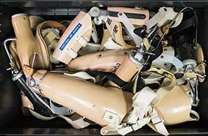
A drawer full of upper extremity prosthetics demonstrates the diversity of the patients treated at the U-M Orthotics & Prosthetics Center. (Image: Austin Thomason, Michigan Photography.)
Editor’s Note: This article is written by Allison Willson on Sept. 17, 2015. This story was reposted courtesy of Medicine at Michigan magazine.
A paradigm shift in prosthetics is happening at the University of Michigan (U-M).
Scientists have developed surgical techniques and technology that will allow amputees to control prosthetic limbs just as they would their own arms and legs.
They have advanced the field of prosthetics through solutions grounded in human biology. Their truly novel work has the potential to restore fine motor control and sensation to people who have lost limbs, so that they might one day be able to tie their shoes or hold and feel the hands of their loved ones.
“We’ve come a long way because of the team we have,” said reconstructive and plastic surgeon Paul Cederna, MD ’89, of a longstanding collaboration between U-M engineers, surgeons, clinicians, research scientists and other experts.
“The engineers have this incredible potential to design all sorts of things,” he said. “The clinicians have a million clinical problems to address, but they don’t have the skillset to conceptualize and design a solution. Getting the two together makes our work incredibly efficient and incredibly fruitful in bringing our big ideas to patients.”
Not surprisingly, it was a challenge put forth by a demanding benefactor that set the U-M team on course toward this elegant solution.

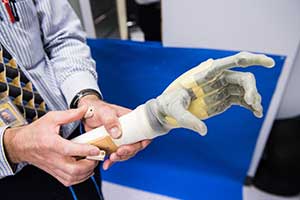
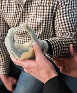
Patients might one day be able to tie their shoes or hold and feel the hands of their loved ones. (Image: Austin Thomason, Michigan Photography.)
Back in 2006, a team of U-M researchers met with the Army Research Office to address this challenge. The Army Research Office had recently given the team $4.5 million based on a grant proposal. The proposal went on for hundreds of pages about the negative effects of “stuffing nails” —or electrodes, more accurately—into nerves, something the Army Research Office is vehemently against. The grant was part of the Multidisciplinary University Research Initiative (MURI) whose purpose was to create “the next generation of artificial limbs.”
Cederna was part of that team, which was focused on developing an interface that would allow amputees to control robotic arms using the motor signals carried by their remaining peripheral nerves—enabling the nerves that once flexed fingers, for example, to flex prosthetic fingers. Cederna first heard about the project through a colleague he had known since his years as an engineering undergrad at U-M, Daryl Kipke, PhD, a U-M professor of biomedical engineering from 2001-13.
“He was a brain guy, and he was putting these really amazing, high-tech electrodes into the brain to record signals,” said Cederna, who is now the Robert Oneal Collegiate Professor and chief of plastic surgery and a professor of biomedical engineering. “I was a nerve guy. I said, ‘How about I take your brain electrodes, and we put them into peripheral nerves to control prostheses?’”
However, that entire premise was out the window when the team learned during that heated kickoff meeting with the Army Research Office.
“I didn’t give you millions of dollars to put electrodes into nerves,” said Cederna remembering what the project manager told them. “I gave you millions of dollars because I believe in this group, and I think you can come up with something better.”
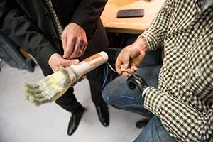
Jeff Wensman, U-M Orthotics & Prosthetics Center clinical/technical director, demonstrates how electrodes placed on the surface of the skin can be used to control prosthetics. (Image: Austin Thomason, Michigan Photography.)
What nerves want
Later during the MURI site team visit, Cederna was having lunch with Melanie Urbanchek, PhD, a muscle physiologist and research associate professor of plastic surgery, and David C. Martin, PhD, who was then a U-M professor of materials science, macromolecular science, and biomedical engineering, and the team’s principal investigator. Eager to fill the drawing board with a more nerve-friendly approach to their research, they considered what the severed nerves would naturally “want.” Being a “nerve guy,” Cederna knew the answer: Nerves want a neuromuscular junction.
The nerves in an amputee’s residual limb continue to grow and send motor signals to their missing arm or leg. But with no muscle to grow into, or innervate, they gnarl into a tangled mess called a neuroma, which can often be painful for the amputee.
“We came up with the idea of putting small pieces of muscle on the end of the nerves, so that the nerve would grow back into that muscle and reinnervate it,” Cederna said. “Then we could use that muscle as an interface to control the prosthesis.”
They called it a regenerative peripheral nerve interface (RPNI). Originally, their plan was to coat the small pieces of muscle with an organic electro-conductive polymer before attaching them to a nerve responsible for a motor function. So when the amputee thinks about moving their thumb, for example, the muscle at the end of the nerve that controls thumb movement would fire a signal. Polymer would pick up that signal and convert it to an electronic signal, which would wirelessly control a motor to move the thumb on the prosthesis.
The approach was novel because the majority of surgeons and engineers researching nerve interfaces were—and still are—placing electrodes on the surface of the skin or the nerve, suturing small wires into the nerve or penetrating the nerve with needles. This is a short-term solution because the nerves are delicate and build scar tissue over time. With the U-M team’s RPNI, that would not be an issue.
Early on, the team planned to culture small pieces of muscle for the interface in the neuromuscular lab, using a tissue engineering approach to grow the muscle cells. It worked well enough, but it was complicated and time-consuming. It also would create FDA approval hurdles down the line. Urbanchek knew there was a better way.
“I was the one growing the muscle cells, and I became rather tired of growing those cells,” Urbanchek said. “There are just so many variables to keep those cells alive, and I thought, ‘Why don’t we just transfer a whole muscle from someplace else? We already know that works.’”
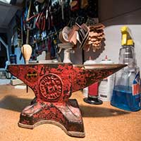
U-M Orthotics & Prosthetics Center staff combine this old-school tool with groundbreaking technology to create prosthetics for patients. (Image: Austin Thomason, Michigan Photography.)
Signals firing
They knew it worked because of research done decades ago at U-M by Bruce Carlson, MD/PhD, former chair of the Department of Anatomy and Cell Biology, now the Department of Cell and Developmental Biology, and professor emeritus of anatomy.
Carlson was interested in the physiology of muscle regeneration. So in the late 1980s, he and John Faulkner, PhD, professor emeritus of molecular and integrative physiology and of biomedical engineering, devised a cross-age transplantation experiment in which entire muscles from older rats were grafted into young rats and vice versa. They were trying to determine whether the young muscle would regenerate better than the old muscle in the older host.
During the transplants, Carlson imbedded the stump of the nerve into the muscle. What he found was that it was not the age or other characteristics of the muscle that determined whether it would regenerate. It was the environment, and nerve reinnervation (restoration of the nerve function) was a major factor in the success or lack of success.
Once more, with feeling
Applying Carlson and Faulkner’s technique to create the RPNIs, Cederna performs microsurgery to separate the individual nerve fascicles—or small bundles of fibers—that remain after an amputation. He then transplants 3 x 1 cm pieces of muscle onto the end of each divided nerve. Reinnervation happens quickly. Urbanchek said it is common to see signals firing after one month, and at three months, the signals are mature enough to control a prosthesis.
“Moving from cell culture to whole muscle made things incredibly simplistic,” Cederna said. “It also made our work scalable to the average surgeon in the community who might do this one day because the average surgeon is not doing cell cultures. So to take a little piece of muscle and transfer it onto the nerve was a huge deal.”
U-M Orthotics & Prosthetics Center staff use these tools and devices to create prosthetics for patients. (Image: Austin Thomason, Michigan Photography.)
A new sensation
After about 350 animal experiments with the RPNIs, the team realized that none of the rats were getting the painful neuromas that often plague patients who have limb loss.
Of the 185,000 amputations each year and 2 million people living with limb loss in the U.S. alone, every one of those amputees will get a neuroma. Just under half of them will have neuromas that cause them significant pain.
“It’s a burning sensation, and it burns almost 24 hours a day, seven days a week,” said Mike Moran, a patient of Cederna’s who lost his arm on the job as a professional painter, when humidity in the air caused his ladder to come in contact with the electricity from a power line. “Really the neuroma pain was greater than all of the psychological trauma, the spiritual trauma and the physical trauma of losing my arm.”
Moran has had two surgeries to treat his neuroma, during which Cederna cut the nerves. Both procedures alleviated his pain, but only until the nerves grew back. Cederna has since transitioned his muscle graft surgery into the clinic. Though Moran has generally been hesitant to have another operation—after enduring a total of 25 surgeries over the years to treat his injuries—he has expressed interest in the new procedure, as he still grapples with neuroma pain 15 years after his accident.
“With typical neuroma patients, 30-40% get better with the current treatments,” Cederna said. “But we found the muscle graft surgery tends to make the pain a lot better or go away completely. So we started doing it for the clinical purposes of controlling neuromas and phantom limb pain [the sensation of the presence of an amputated limb].”
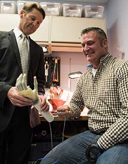
Cederna explains how nerve signals and electrodes will be used to control prosthetics to his patient, Mike Moran. (Image: Austin Thomason, Michigan Photography.)
Patients, please
Moving forward with the muscle graft surgery for clinical purposes was a major breakthrough for the team. It positioned them to do the procedure on human patients, who would then have the first phase of the RPNI in place for future clinical trials. Cederna has successfully performed the surgery on more than 20 patients.
Another breakthrough came when the team discovered they could utilize nerve signals for prosthetic control without the electro-conductive polymer. Cederna said they have currently stopped working with the polymer due to regulatory hurdles preventing its use in human patients, but there is a possibility they may incorporate it into the RPNI down the line.
For now, as the team found, the attached pieces of muscle amplify the nerve signals enough so that they can be recorded by a small electrode placed on the muscle. The team uses the data from those nerve recordings to program algorithms into the electronics of a robotic hand to tell it how to respond to particular signals. This would not be possible without the muscle amplification of the signal.
“It’s fundamentally hard to record from the nerve directly because the signals are so small,” said Cindy Chestek, PhD, an assistant professor of biomedical engineering and the team’s expert in brain-machine interfacing.
“Now, we can record from the muscle and get a much bigger signal from the nerve and because Paul dissects the nerve really well, it’s a much more specific signal. You really want to be recording from smaller and smaller groups of neurons, even individual neurons, in the nerve if you want to control different movements at the same time, for the fine motor control. You can figure out which grasp the subject is trying to make or work toward continuous position control, and all of these things are just not available in existing prosthetic limbs.”
Making those things available will likely require more advanced electrode technology, said Nicholas Langhals, PhD, a U-M research assistant professor of plastic surgery, who has contributed his expertise in signal processing to the effort. He is collaborating with colleagues in electrical engineering for pilot studies to develop flexible multichannel electrodes to wrap around the individual RPNIs. This will potentially allow the team to record smaller signals from more locations on the muscle and get more data for fine motor control.
The sensors on the prosthesis could pick up tactile information. (Image: Austin Thomason, Michigan Photography.)
The next generation
The team is also working to develop an implantable device that would connect to the RPNIs to pick up the nerve signals and feed them back to the prosthesis. Cederna said it will be similar to a pacemaker box—only a complicated pacemaker box.
“It’s going to have to be a lot more powerful from a computer technology standpoint,” Cederna said, “because it has so much more information that it needs to understand and interpret.”
That information also could include sensory feedback if the device were connected to a sophisticated enough prosthetic limb with built-in electronic sensors. The sensors on the prosthesis could pick up tactile information like pressure, temperature and texture, and the electrodes on the RPNIs could, in theory, receive those signals and feed them back to the amputee.
“Getting the control signals down to the motors on the prosthesis is relatively straightforward at this point,” said Brent Gillespie, PhD, a U-M associate professor of mechanical engineering whose Haptix Lab studies tactile sensation in human-machine interface devices, including prosthetics.
“We’ve shown it in monkeys and rats, and we’re hoping to move into humans pretty soon. But getting sensory feedback from the electronic sensors back into the peripheral nervous system is new and relatively unexplored. It’s hard to ask the rat or the monkey what it feels, so we’re basically going to need to do this exploratory research in humans.”
“The more [your prosthetic limb] moves realistically and the more sensation it provides, the more it will be incorporated into your body image,” Cederna said. (Image: Austin Thomason, Michigan Photography.)
The clinical trials to test the function of the RPNI for motor control and sensory feedback in human patients are expected to begin within the year, and ideally the first patients would be enrolled by this summer.
The team is looking to private foundations and public money for funding, and they expect patients to be able to use the RPNI system, which will be compatible with any type of prosthesis, at home in the next few years. They are confident it will be superior to any of the current approaches to prosthetic control, which lack high-fidelity movement and sensory feedback.
The team is well on its way to enabling that “next generation of artificial limbs” MURI challenged them to create back in 2006. Although, “artificial” may be a less accurate description of what this new generation promises.
“We want to get people as close as possible to where they were before they had their horrible injury or cancer operation or whatever it was that led to limb loss,” Cederna said.“The more [your prosthetic limb] moves realistically and the more sensation it provides, the more it will be incorporated into your body image. The more it’s incorporated into your body image, the more likely you are to start using it like a natural limb—to make gestures or to hold your child’s hand … I see the patients, and I know what they’re going through, and I know what we’re doing is really going to have an impact.
“I feel this. I really feel it.”
University of Michigan
www.umich.edu
U-M Orthotics & Prosthetics Center
www.uofmhealth.org


