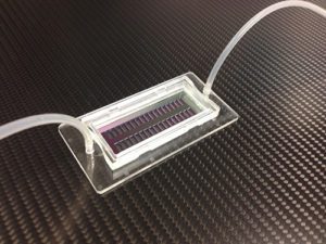
The new microhole chip can be populated with 200,000 single cells, each held in place in separate holes. [Image from Fraunhofer IBMT]
Traditional fluorescence-activated cell sorting (FACS) gives an estimate of the number of tumor cells in a patient’s bloodstream. If there is a higher concentration of tumor cells, there is a greater chance that they will increase metastasis. However, FACS is limited to a small set of colored dyes that can be used.
“In FACS, the tumor cells are stained with fluorescent dyes and sorted into separate collection vessels,” said Thomas Velten, lead researcher of the Fraunhofer Institute for Biomedical Engineering IBMT team that created the chip, in a press release. “Eventually you run out of colors that are sufficiently contrasting to be able to tell them apart. Moreover, there are no suitable markers available for certain types of tumor cells, which are therefore not detectable by FACS analysis.”
FACS analyses also can’t be narrowed down to a single cell because the collection vessel has thousands of cells. But the new microhole chip can pick out single cells from a blood sample and place them on separate holes to analyze.
“It’s easy to select cells because each one has its own specific position in the array where they are lined up like ducks in a row. Each cell is placed on a hole but cannot slip through it. A slight under pressure is applied to the cells that hold each one in its allotted place by suction,” said Velten.
The researchers selected suspicious-looking cells using a microscope and those cells were analyzed using Raman spectroscopy which exposes the cells to light within a specific frequency range. Tumor cells were able to scatter light to make them clearly identifiable. This method is limited because it can’t be used on conventional arrays with glass or polymer substrates, but the new IBMT chip can.
The microhole chip can also hold up to 200,000 cells, organizing them into separate holes for each cell within minutes. Micropipettes remove tumor cells from the chip for analyzing.
“This is important because the bloodstream contains only a small quantity of circulating tumor cells, which cannot be detected unless the blood sample is large enough. Older lab-on-chip technologies cannot accommodate more than 1,000 cells, which is too few for this kind of application,” said Velten.
Researchers suggest that the microhole chip could be used as a selection system for protein-producing cells like the ones that make insulin and other biopharmaceuticals. They can also be used for in-vitro modeling of physiological barriers like blood-brain barrier if micropores are well-defined.
[Want to stay more on top of MDO content? Subscribe to our weekly e-newsletter.]


Very excited to get this information, and I am starting to design and fabricate microfluidc devices for cell culture and cell capture. And I would like to get more valuble advice from you, because I am just beginning with this issue. Thanks, anything you talk to me may be good, do not hesitate to tell me.
Regards,
Hong Jiang
Thanks for the comment!