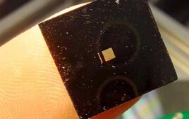
Graduate student Seongjun Park holds an example of a new flexible fiber, which is no bigger than a human hair and has successfully delivered a combination of optical, electrical, and chemical signals back and forth into the brain. [Image from Young Gyu Yoon/MIT]
The fibers replicate the softness and flexibility of brain tissue which allows for brain implants to remain and retain their functions over longer periods than with stiff, metallic fibers. The method also allows for more data collection.
In lab tests with mice, the researchers could inject viral vectors that carried genes that sensitive neurons to light, called opsins, through one of two fluid channels in the flexible fiber. Once the opsins took effect, the researchers sent a pulse of light through the optical waveguide in the center. They recorded the neuronal activity that happened as a result using six electrodes that could identify different reactions – all through a single flexible fiber that was 200 micrometers wide.
Separate devices like needles to inject viral vectors for optogenetics, optical fibers for light delivery and electrodes for recording have been the focus of other research efforts.
“We said, wouldn’t it be nice if we had a device that could just do it all,” Polina Anikeeva, professor in the departments of materials science and engineering and electrical engineering and computer science at MIT, said in a press release. ““It can deliver the virus [containing the opsins] straight to the cell, and then stimulate the response and record the activity — and [the fiber] is sufficiently small and biocompatible so it can be kept in for a long time.”
The fibers have the potential to be used to observe activity coming from varying regions of the brain, according to the researchers. In some of the first tests, the researchers put probes in two different regions of the brain at one time and measured how long it took for responses to be sent from each region to the other.
The fiber is able to work through conductive wires that could hold the flexibility needed while carrying electrical signals as well. A composite of conductive polyethylene mixed with graphite flakes was used to make the fiber. Polyethylene was formed into layers and sprinkled with graphite flakes and then compressed and more compressed polyethylene layers sprinkled with graphite flakes were added. Using the layering method increased the conductivity of the polymer by four or five.
After 11 days, the specific sensitizing agent that the researchers used was able to create a noticeable effect. They hope to make the fibers thinner in the future to make them replicate neural tissue even more.
“The authors report some remarkably sophisticated designs and capabilities in multifunctional fiber devices, where they create a single platform for colocalized expression, recording, and illumination in optogenetics studies of brain function,” said John Rogers, professor of materials science and engineering, biomedical engineering and neurological surgery at Northwestern University. “These types of advances in technologies and tools are essential to progress in neuroscience research.”
The research was published in the journal Nature Neuroscience and was supported by the National Institute of Neurological Disorders and Stroke, the National Science Foundation, the MIT Center for Materials Science and Engineering, the Center for Sensorimotor Neural Engineering and the McGovern Institute for Brain Research.