Doctors at the University of Connecticut are using 3D printing technology to help them practice some delicate brain surgery.
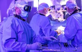
Since 2012, physicians have been using a procedure to treat strokes called a mechanical thrombectomy, guiding a catheter through a patient’s arteries and vessels into the brain to remove blood clots.
Medical physicist David Brotmann came up with the idea of creating an exact model of those blood vessels by converting medical scans into an image that could recognized by a 3-D printer.
Doctors learning the procedure could then practice on the plastic model, threading a wire through it and up to a piece of cotton or other substance that simulates a clot. The more they practice, the less likely something will go wrong during the actual surgery.
“The way previously to practice this technique was on a living patient,” says Brotman. “I don’t think you want that to be you or me.”
Brotman brought his idea to Dr. Clifford Yang, a cardiac radiologist at UConn Health with an electrical engineering background, who in turn showed it to Dr. Charan Singh, an interventional radiologist who teaches the thombectomy procedure at UConn.
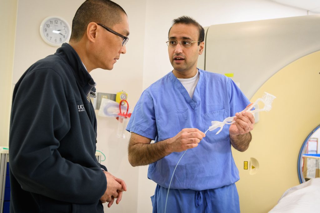
Dr. Charan K. Singh, right, holds a 3-D printed model of arteries and a catheter while speaking with Dr. Clifford Yang at UConn Health. (Peter Morenus/UConn Photo)
Together they figured out how to translate the medical scans into printable images.
“The scans, either CT or MRI, give you a bunch of slabs, layer by layer of what the vessels look like,” says Yang. “We are able to transfer that to files the 3D printer can recognize and take those slabs and print layer by layer over several hours.”
Singh said using the model is a much more cost-effective way to teach the procedure to residents than using a two-dimensional computer simulation and much safer than doing it for the first time on an actual patient. The printed model costs about $14, he says.
UConn currently has one set of images it uses to create models, which come from an anonymous patient. Eventually, the doctors say they will be able to convert medical scans almost immediately, allowing neurosurgeons and others to practice on a model of any patient’s unique vascular structure before going into surgery.
“Everybody’s anatomy is different,” says Singh. “So, in a tough case, it might be easier or better to practice outside the body, even one time, before going into a more serious situation.”
UConn is making its first-of-its-kind 3D image files available to any hospital that wants to create their own models.
Brotman, who now works at Yale, says he thought about applying for intellectual property protection for his idea. But he said it was more important to get the technology out there than to make money from it.
Dr. Justin Caplan, who teaches neurosurgery at John’s Hopkins, says there are similar mock-up devices on the market, including some that can simulate blood flow, but those are expensive. He said as the technology has gotten cheaper, using 3D printing in neurosurgery is an idea that is catching on nationally.
“Strokes and other emergencies, you typically can’t practice on an individualized patient ahead of time,” he says. “But for elective procedures, such as aneurisms, you could custom 3-D print that exact patient’s exact aneurism and practice. There’s a benefit there for the trainee and for a surgeon in tricky cases.”
Brotman said his biggest hope is that 3D-printed models will provide a cheap way to teach doctors and medical students in rural areas, third-world countries, or others without access to high-tech surgical simulators.
“A doctor can even take this home and practice, it’s a real paradigm change,” he says.