[Image courtesy of ETH Zurich]
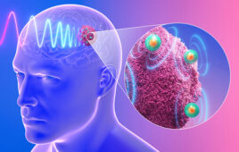
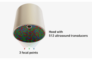
The research team designed a helmet-shaped device equipped with several hundred ultrasound transducers that emit short, low-intensity pulses. These pulses interfere with each other inside the brain, forming distinct focal points similar to the way light waves create a hologram. In laboratory experiments on mice, the system produced three to five simultaneous stimulation points with precise targeting.
Researchers believe the method could inform future treatments for neurological disorders such as Alzheimer’s disease, Parkinson’s and depression. The research was published in the Nature Biomedical Engineering journal.
“Given that the brain operates in networks, it’s easier to activate or inhibit a brain network if you stimulate it at multiple points simultaneously,” said Daniel Razansky, a professor at ETH Zurich and the University of Zurich, who led the study in collaboration with colleagues at New York University.
How the ultrasound holograms work
Neuromodulation is performed through the skull without the need for surgery. In lab experiments, a mouse’s head was positioned inside a hood-shaped array containing hundreds of transducers, controlled by electronics that generated brief ultrasound pulses. The waves combine inside the brain to create the multiple focal points of stimulation. The system, about 8cm in diameter, can modulate activity in brain networks while using less ultrasound intensity than single-spot methods.
“The less intense the ultrasound, the safer this process is for the brain,” said Razansky.
Earlier approaches to the method suffered from an all-or-nothing effect, providing too little intensity with no impact. At the same time, higher power risked overheating tissue or causing uncontrolled activation of large brain regions. Strong ultrasound exposure can also damage blood vessels or cause unwanted heating of the brain or skull tissue.
The researchers believe that low-intensity focused ultrasound can act through brief temperature increases and mechanical effects on ion channel proteins located on neuron surfaces. These proteins regulate ion flow into and out of cells to help control electrical signaling in the brain. The researchers want to study the method further to understand exactly how ultrasound pulses activate or inhibit neurons.
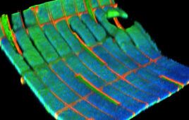
According to the team of researchers, the new ultrasound system can modulate brain networks and visualize their activation in real-time, allowing researchers to see which areas respond to stimulation.
What’s next?
The U.S. National Institutes of Health primarily funded the project and collaboration with New York University. Razansky said current funding restrictions on international partners will limit further work under the same program, but his group plans to continue with other funding sources.
Now, the researchers plan to test the technology in animal models of neurological diseases, including Alzheimer’s, epilepsy, tremor, Parkinson’s and stroke recovery.
“We rely on animals for our research,” Razansky said. “It won’t be possible to research these developments at such an early stage in humans. We first need to learn how to control the intervention and ensure that it is safe and effective for the treatment of brain diseases.”
Razansky’s group at ETH Zurich specializes in ultrasound and optical imaging technologies, focusing on system design, experimental methods and data analysis.