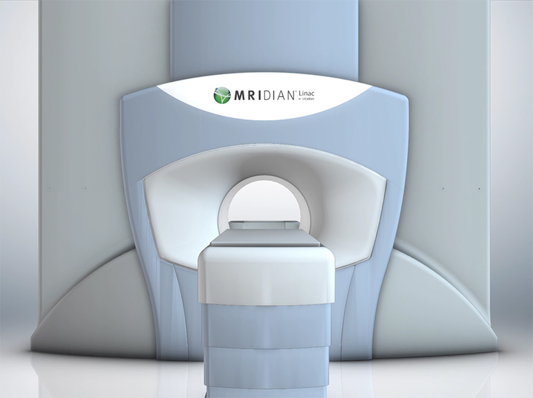
ViewRay, Inc. announced that the company received 510(k) clearance from the U.S. Food and Drug Administration (FDA) to market the MRIdian Linac system, the company’s next generation linear accelerator-based MRI-guided radiation therapy system.
“FDA clearance of the MRIdian Linac is a transformative milestone for ViewRay,” says Chris A. Raanes, president and chief executive officer of ViewRay. “We believe that availability of the world’s first commercial system to combine MRI for soft-tissue visualization and a compact linear accelerator will lead to a new standard of care in radiation oncology. Clinical experience with ViewRay’s first generation MRIdian System has demonstrated the benefits of on-table adaptive therapy and real-time MRI guidance, as clinicians are seeing for the first time how much tumors and organs move and change shape during the course of treatment.”

(Image credit: ViewRay)
The first two MRIdian Linac systems in the United States are expected to be installed at Henry Ford Hospital in Detroit and Barnes-Jewish Hospital at Washington University in St. Louis.
“Radiation oncologists have long awaited the availability of a clinical system that integrates MR-imaging with linac radiation delivery, as this is a major breakthrough in improving the efficacy of radiation therapy for patients,” says Benjamin Movsas, MD, chairman of Radiation Oncology at the Henry Ford Cancer Institute in Michigan. “At the Henry Ford Cancer Institute, we’re proud to be among the first centers to offer treatment with MRIdian Linac. The ability to see what’s happening inside the body while the patient is undergoing radiation therapy and ensure the radiation continually remains on target is a huge leap forward and will help us to improve patient outcomes.”
“With MRI-guided radiation therapy, we’re able to watch the movement of tumors and organs in the body as radiation is being delivered and adapt the dose of radiation in real-time, to help ensure the maximum dose reaches the tumor and that surrounding healthy tissue is spared,” says Sasa Mutic, PhD, director of Radiation Oncology Physics at Washington University School of Medicine. “This technology helps us treat tumors such those in the lung, liver and pancreas where increased precision is important due to nearby organs and other critical structures.”


