[Image from NYU]
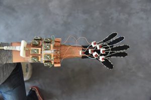
The NYU study showcased how a MRI element design that is woven into garment-like detectors can create high-quality images of moving joints. The researchers suggest that the glove prototype can be useful for diagnosing repetitive strain injuries like carpal tunnel.
The MRI glove shows how different tissue types impact each other as they naturally move, which can also give more insight into how the hand works while guiding surgery with hand images in a more realistic position and designing better prosthetics.
“Our results represent the first demonstration of an MRI technology that is both flexible and sensitive enough to capture the complexity of soft-tissue mechanics in the hand,” said lead author Bei Zhang, research scientist at the Center for Advanced Imaging Innovation and Research (CAI2R), in a press release.
MRI machines exposes tissues to a magnetic field that allows hydrogen atoms to attach to and create an average magnetic force in one direction with each tissue slice. The magnets can then be moved out of equilibrium by waves of electromagnetic force where they begin to spin like tops and emit radio signals, which indicates where they are located that can be translated into images.
An MRI also depends on the ability of radiofrequency coils to convert radio waves into an electrical current. Radio waves don’t produce as many electrical currents inside received coils, so they end up creating their own magnetic fields, preventing nearby coils from capturing a clean signal.
The researchers wanted to create a structure with high impedance that blocks current and measures how hard the force in any magnetic waves is pushing voltage as it tries to create a current. The glove-shaped MRI component receiver coils no longer create magnetic fields that interfere with other nearby receivers. It creates images of freely moving muscles, tendons and ligaments in a hand as it performs tasks like playing piano and grabbing objects without the need for rigid structures.
Hydrogen atoms make up an MRI signal which means the technology is able to image soft tissue structures that are rich in water. This is why MRI is ideal for imaging muscles, nerves and cartilage that can be typically harder to study with other non-invasive methods. However, tendons and ligaments are made of dense proteins that show up as black bands on the sides of bones. With the glove, the coils showed how the black bands moved with the bones.
“We wanted to try our new elements in an application that could never be done with traditional coils, and settled on an attempt to capture images with a glove,” said senior author Martijn Cloos, assistant professor from the CAI2R institute in the Department of Radiology at NYU Langone Health. “We hope that this result ushers in a new era of MRI design, perhaps including flexible sleeve arrays around injured knees, or comfy beanies to study the developing brains of newborns.”
The research was published in the journal Nature Biomedical Engineering.