A team of biomedical engineers at Washington University in St. Louis, led by Lihong Wang, PhD, the Gene K. Beare Distinguished Professor of Biomedical Engineering, has developed the world’s fastest receive-only 2-D camera, a device that can capture events up to 100 billion frames per second.
That’s orders of magnitude faster than any current receive-only ultrafast imaging techniques, which are limited by on-chip storage and electronic readout speed to operations of about 10 million frames per second.
Using the Washington University technique, called compressed ultrafast photography (CUP), Wang and his colleagues have made movies of the images they took with single laser shots of four physical phenomena: laser pulse reflection, refraction, faster-than light propagation of what is called non-information, and photon racing in two media. While it’s no day at the races, the images are entertaining, awe-inspiring and represent the opening of new vistas of scientific exploration.
The research appears in the Dec. 4, 2014, issue of Nature.
“For the first time, humans can see light pulses on the fly,” Wang says. “Because this technique advances the imaging frame rate by orders of magnitude, we now enter a new regime to open up new visions. Each new technique, especially one of a quantum leap forward, is always followed a number of new discoveries. It’s our hope that CUP will enable new discoveries in science — ones that we can’t even anticipate yet.”
This camera doesn’t look like a Kodak or Cannon; rather, it is a series of devices envisioned to work with high-powered microscopes and telescopes to capture dynamic natural and physical phenomena. Once the raw data are acquired, the actual images are formed on a personal computer; the technology is known as computational imaging.
The development of the technology was funded by two grants from the National Institutes of Health that support pioneering and potentially transformative approaches to major challenges in biomedical research.
 “This is an exciting advance and the type of groundbreaking work that these high-risk NIH awards are designed to support,” said Richard Conroy, PhD, program director of optical imaging at the National Institute of Biomedical Imaging and Bioengineering, part of the NIH. “These ultrafast cameras have the potential to greatly enhance our understanding of very fast biological interactions and chemical processes and allow us to build better models of complex, dynamical systems.”
An immediate application is in biomedicine. One of the movies shows a green excitation light pulsing toward fluorescent molecules on the right where the green converts to red, which is the fluorescence. By tracking this, the researchers can get a single shot assessment of the fluorescence lifetime, which can be used to detect diseases or reflect cellular environmental conditions like pH or oxygen pressure.
Wang envisions applications in astronomy and forensics, where the advanced imaging frame rate could analyze the temporal activities of a supernova that occurred light years away, or track and predict the movements of thousands of potentially hazardous pieces of “space junk,” refuse of old satellites and jettisoned space craft hurtling about at high speed in outer space. In forensics, CUP might be used in reproducing bullet pathways, which could once again open up the Kennedy assassination conspiracy theories and revive a more accurate analysis of the strange physics of the “magic bullet.”
Wang and his collaborators essentially added components and used algorithms to complement an existing technology known as a streak camera, which measures the intensity variation in a pulse of light with time. While a streak camera is fast, it gives only a one-dimensional view, which “is not intuitive — much analogous to watching a horse race through a distant vertical slit,” Wang said. “We expanded the view into 2-D space, more like what we see in the real world.”
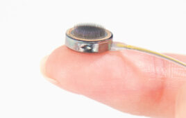
CUP photographs an object with a specialty camera lens, which takes the photons from the object on a journey through a tube-like structure to a marvelous tiny apparatus called a digital micromirror device (DMD), smaller than a dime though hosting about 1 million micromirrors, each one just seven by seven microns squared. There, micromirrors are used to encode the image, then reflect the photons to a beam splitter which shoots the photons to the widened slit of a streak camera. The photons are converted to electrons, which are then sheared with the use of two electrodes, converting time to space. The electrodes apply a voltage that ramps from high to low, so the electrons will arrive at different times and land at different vertical positions. An instrument called a charge-coupled device (CCD) stores all the raw data. All of this occurs at the breathtaking pace of 5 nanoseconds. One nanosecond is a billionth of a second.
Wang’s work with CUP pushes the dimensional limits of fundamental physics and also pushes the limits of deep imaging of biological tissues, one of Wang’s research specialties.
“Fluorescence is an important aspect of biological technologies,” he says. “We can use CUP to image the lifetimes of various fluorophores, including fluorescent proteins, at light speed.”
In the astronomy world, CUP can be a game-changer, Wang says.
“Combine CUP imaging with the Hubble Telescope, and we will have both the sharpest spatial resolution of the Hubble and the highest temporal solution with CUP,” he says. “That combination is bound to discover new science.”