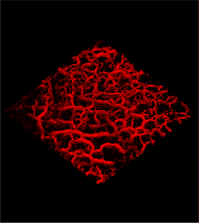
(Credit: VivoSight)
Skin cancer can now be detected in under 30 seconds.
European researchers have developed a scanner that uses an infrared laser beam to detect blood vessels grown by malignant melanoma.
Traditional steps for diagnosis include a visit to a dermatologist, waiting for laboratory results, undergoing a biopsy, and potentially additional tests.
(Credit: VivoSight)
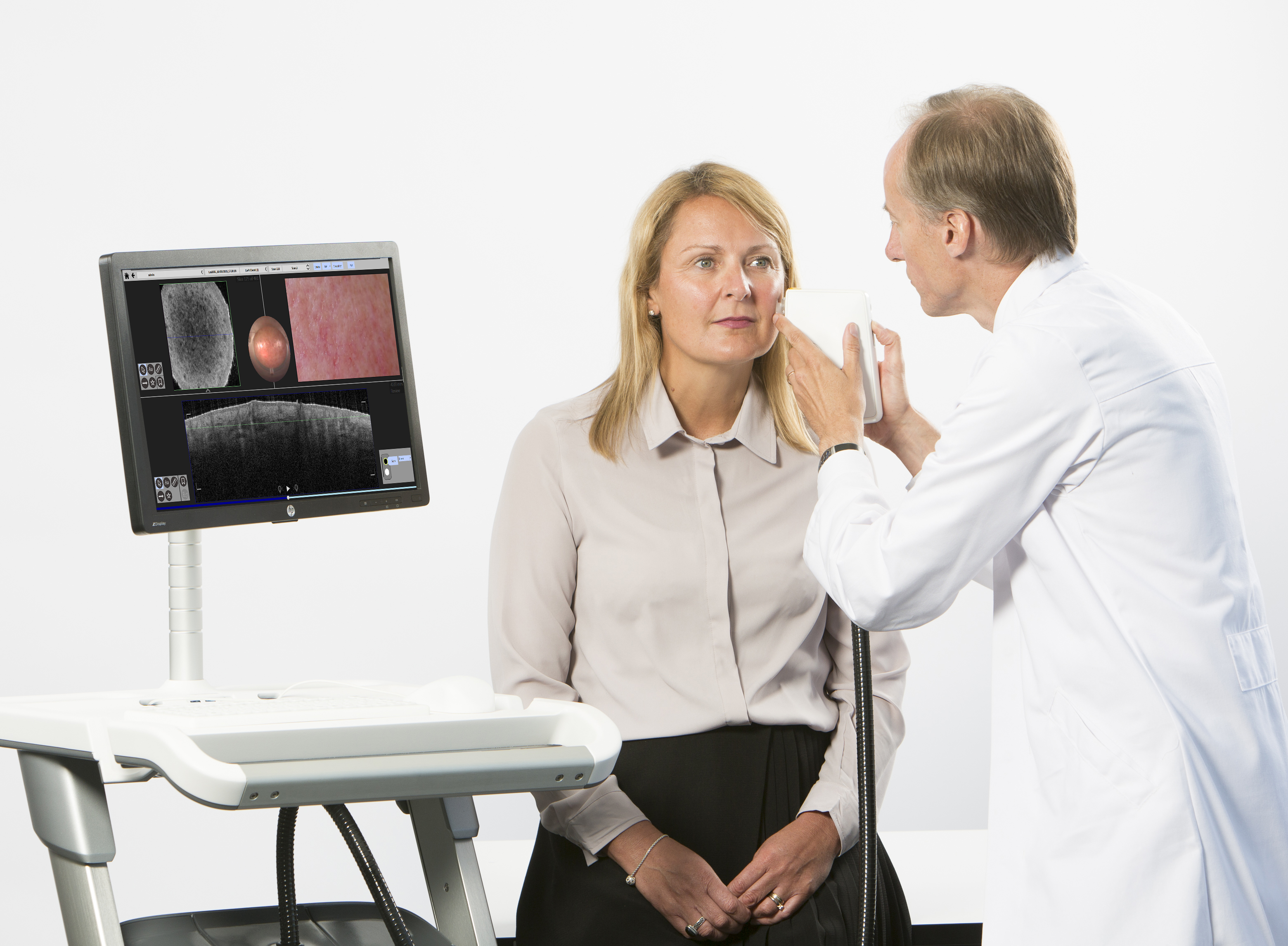
The device is a handheld laser, VivoSight, used to see 1mm underneath the skin. It creates a 3D color image of the blood vessels. It uses Optical Coherence Tomography (OCT).
It takes four frames per second and complies them so that physicians may see if something has moved from frame to frame.
“Since melanomas need oxygen to grow and survive, they grow their own blood vessels,” says Jon Holmes, Chief Executive and Chief Technology Officer of VivoSight.
“As the cancer develops and becomes more malignant, they become increasingly distorted and malformed, differing in appearance from healthy vessels.”
Prior to the development of this device, the ability to detect and see the vessels in a suspicious lesion in real time had not been possible.
“Using D-OCT we can see movement of blood against the solid tissue structures, something we have never been able to do before in a clinical setting. It’s like looking out at night and seeing cars’ headlights flowing along a motorway, only at depths of nanometers under the skin,” says Holmes. “But it appears cancers don’t take the direct route! Their vessels are like twisty, branching country lanes that get narrower and wider. Our clinical team thinks that these ‘shapes’ are key to understanding the cancer. Our scanner shows these vessels in gorgeous detail.”