A recent case letter in the current issue of the Journal of Oral Implantology has described a technique to create and position titanium mesh to improve guided bone regeneration.
In collaboration, Loma Linda University, King Khalid University, and King Saud University, doctors encountered a patient who had several missing front teeth due to an injury that resulted in a large gap referred to as a bony defect. The authors began with a bone graft, followed by dental implants that would support the replacement teeth. They created a virtual 3D model of the patient’s mouth and bony defect, then, with a computer program, determined the correct position and placement of the bone graft based on the desired location of replacement teeth. They 3D printed a model and fabricated the titanium mesh to fit the intended bone position and volume. The authors filled the titanium mesh with bone harvested from a remote location in the patient’s mouth and secured it in the site of the missing teeth with fixation screws.
By digitally creating the titanium mesh, the authors were able to ensure the material was the precise shape and volume needed for the patient’s bone defect. This reduced the chance that the titanium mesh would cause an unfavorable outcome during healing, including mesh exposure and irritation. During surgery, the authors ensured proper placement of the titanium mesh and bone graft with a positioning jig fabricated from a clear plastic mold that used the existing teeth to hold the mesh in place while it was being secured. Finally, to improve wound healing and bone stability, they applied fibrin membranes rich in platelets.
“Virtual 3D planning has become more popular in recent years when planning for implant placement,” said author Aladdin J. Al-Ardah. “Our technique ensures precise augmentation of the bony defect and an accurate positioning of the mesh.” The authors concluded that prefabrication of titanium mesh also reduces surgical time and ensures that the location of replacement teeth with be correct. Future work is needed to improve fabrication of the positioning jig as well as to determine the dental procedures for which this technique would be appropriate.
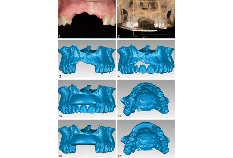
Preoperative frontal intraoral view Credit: Journal of Oral Implantology