A 3D-printed heart made a complex surgery safer for a child patient in Los Angeles.
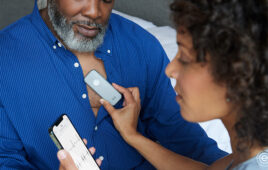
Dr. Richard Kim, a cardiac surgeon at Children’s Hospital Los Angeles, used the printed model to strategize how best to treat Esther Perez, a young patient with a rare type of ventricular septal defect. Perez went in for surgery in November, and remains stable after the operation.
Usually, such planning would require surgery to examine the shape of the anomaly, and decisions would have to be made quickly, while the patient was on the operating table.
Instead, Dr. Jon Detterich, a pediatric cardiologist who specializes in noninvasive cardiac imaging, gathered 2D and 3D MRI scans of Perez’s heart, then used that to build a model a 3D printer could use.
This removed the need for the exploratory surgery and gave Kim more time to plan, making Perez’s procedure – which required complex re-routing of the blood supply – safer.
“Instead of opening the chest and making a decision about how to proceed, I could immediately begin fixing the problem,” said Kim. “A 3D model allowed me to plan the surgery in advance, which meant Esther spent less time in surgery and received less anesthesia – making the procedure safer.”
So far, only a small number of 3D models have been used for heart surgery, so it’s too soon to tell if they improve surgical outcomes,” said Frank Ing, MD, chief of Cardiology and co-director of the Heart Institute at CHLA. “But our experience suggests that using models saves time in the OR – which means increased safety and decreased costs.”
Both Ing and Kim also teach at the Keck School of Medicine of the University of Southern California.