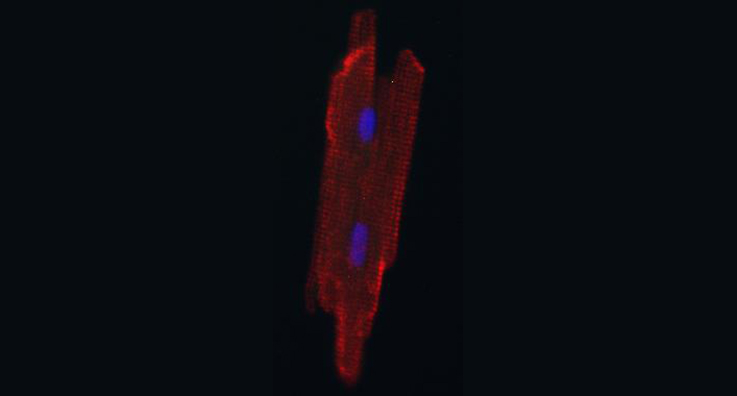
Johns Hopkins researchers grew this adult heart muscle cell inside a newborn rat heart. [Image courtesy of Johns Hopkins Medicine]
Researchers at Johns Hopkins University recently reported that they’ve had success using host animal hearts to grow mature and viable heart muscle cells from stem cells.
Meanwhile, a 1-micron-resolution scaffold made with a 3-D printer and seeded with cells was eventually able to significantly improve heart function when surgically implanted onto the heart of a mouse that had suffered a heart attack, according to the University of Alabama and University of Minnesota researchers behind the study.
The Johns Hopkins researchers think their new cell-growing method could aid heart disease research studies and potentially even lead to new diagnostic tools and treatments.
“Our concept of using a live animal host to enable maturation of cardiomyocytes can be expanded to other areas of stem cell research and really opens up a new avenue to getting stem cells to mature,” said Chulan Kwon, the Johns Hopkins associate professor of medicine who led the study.
The Alabama and Minnesota researchers think their novel heart tissue patch is an advance toward methods to prevent heart failure after heart attacks.
“Thus, the hiPSC-derived cardiac muscle patches produced for this report may represent an important step toward the clinical use of 3-D-printing technology. To our knowledge, this is the first time modulated raster scanning has ever been successfully used to control the fabrication of a tissue-engineered scaffold, and consequently, our results are particularly relevant for applications that require the fibrillar and mesh-like structures present in cardiac tissue,” said the researchers, who were led by Dr. Jianyi “Jay” Zhang of the University of Alabama at Birmingham and Brenda Ogle of the University of Minnesota.
[Want to stay more on top of MDO content? Subscribe to our weekly e-newsletter.]