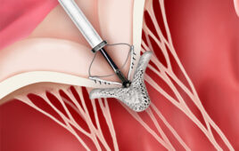
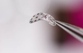
Tremendous strides have been made in tissue engineering and, more specifically, in the development of skin grafts, heart valves, and blood vessels. However, these items are expensive to develop and take time to create. A University of Missouri-Columbia researcher has developed a machine designed to cut down on the time and cost of creating these tissues.
For more than two years, Mark Haidekker, assistant professor of biological engineering, has worked to create a device that examines the quality of grafts and vessels. His goal has been to decrease dramatically the number of possible flaws in the tissues and vessels manufactured. “This is a quality control device that will save lives,” Haidekker says. “This machine increases the success rate of the tissue-engineered items by picking out the rare, but crucial, flaws that may cause serious problems.”
Haidekker describes other methods used to examine tissue as time-consuming and expensive. He says his new device takes a few minutes, not hours, to create 3D images. The device, which involves a technique called optical transillumination tomography, examines tissue using a laser beam and generates a 3D image of the tissue that can be analyzed on a computer. This allows testing of the tissue—for thickness, structure similarity, density, and possible defects—in a non-invasive way.
Information: www.missouri.edu.


