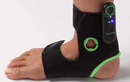
Florian Thürk (left) testing the device [Image from Technische Universität Wien]
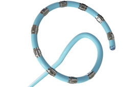
Electrical impedance tomography (EIT) is a new imaging technique created by a collaboration between Technische Universität Wien, Medical University of Vienna and the University of Veterinary Medicine Vienna. The electrode-filled belt is applied directly on the skin for EIT to work. Small currents are passed through the body through the belt and the electrical voltage is measure. The measurements are used to make images of different bodily functions such as lung function.
There is currently no standard method for turning electrical measurements into reliable images, but the research from the three universities has been able to show that the quality of the results given are improved if the evaluation methods were made specifically for each patient. As a result, EIT could be used in intensive care units.
“We have had high hopes for electrical impedance tomography for a number of years,” Stefan Böhme, from the Medical University of Vienna, said in a press release.
The current that goes through the body is a high-frequency current that is weak enough that it can’t be felt by the patient, but is strong enough to be used to calculate the electrical resistance in the body to give a better idea of some of the internal processes of the body.
So far, the belt has shown to be useful for monitoring the functions of the lungs in patients in intensive care. Artificial respiration can cause strain and damage to the lungs if it’s not calibrated to a patient’s specific needs. There are ways to monitor lung function, but most of the time, a patient has to be exposed to multiple X-rays and be transported through a hospital. EIT eliminates the need for that by continuously monitoring lung function directly from a patient’s bed.
“The problem is that there is still no standardized method for determining reliable medical data from the measured results,” said Florian Thürk, a doctoral student in a research group at the Institute of Electrodynamics, Microwave and Circuit Engineering at TU Wien.
“This is considerably more complicated than is the case for computed tomography. From a mathematical perspective, different impedance distributions inside the body can produce identical measurement results. It is difficult to say for sure which of the distributions is actually correct,” Thürk said.
If a little more information is given in the calculation model, more customized evaluation methods could be created.
“High-resolution CT images allow individual parameters – such as the exact location of the contours of the lungs – to be monitored to an impressive degree,” Thürk said. “If we feed this computed tomography data into our evaluation program, we can produce customized evaluation methods that provide much more accurate results than had ever been thought possible.”
The EIT technology has been successfully tested in pigs. EIT results and CT images have shown to be effective when used together. The researchers hope to improve the EIT method to make it the standard method for intensive care units.
“The aim is not to generate the best single frames possible, but rather to derive physiologically relevant parameters from the data in order to directly monitor the lung function. IN the course of their daily work, medical professionals often do not have the time to review individual images – they want the data that is to be monitored to be available to them immediately,” said Eugenijus Kaniusas, one of the researchers on this project.
The research was published in the journal PLoS ONE.