Medical Design and Outsourcing

  • Home
  • Medical Device Business
    • Mergers & Acquisitions
    • Financial
    • Regulatory
  • Applications
    • Cardiovascular
    • Devices
    • Imaging
    • Implantables
    • Medical Equipment
    • Orthopedic
    • Surgical
  • Technologies
    • Supplies and Components Index
    • Contract Manufacturing
    • Components
    • Electronics
    • Extrusions
    • Materials
    • Motion Control
    • Prototyping
    • Pumps
    • Tubing
  • MedTech Resources
    • Medtech Events in 2026
    • The 2026 Medtech Big 100
    • Medical Device Handbook
    • MedTech 100 Index
    • Subscribe to Print Magazine
    • DeviceTalks
    • Digital Editions
    • eBooks
    • Educational Assets
    • Manufacturer Search
    • Podcasts
    • Webinars / Digital Events
    • Whitepapers
    • Voices
    • Views
    • Video
  • 2026 Leadership
    • 2025 Winners
    • 2024 Winners
    • 2023 Winners
    • 2022 Winners
    • 2021 Winners
  • Women in Medtech
  • Advertisers
    • Advertise
    • PR Submissions
    • Eblast Submissions
  • Subscribe

MIT researchers say ‘pencil beam’ lasers improve brain imaging

June 7, 2026 By Sean Whooley

MIT Pencil Beam imaging technology

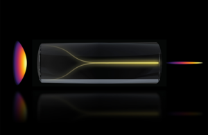
Under the right conditions, a chaotic mess of laser light can spontaneously self-organize into a highly focused “pencil beam.” This schematic shows the pencil beam formation mechanism. [Image courtesy of MIT/the researchers]

MIT researchers say they’ve found a way to enable faster, higher-resolution bioimaging compared to existing technology.

Using a “chaotic mess of laser light,” as MIT put it, can spontaneously self-organize under the right conditions into what the researchers call a “pencil beam.”

Using this method, the researchers acquired 3D images of the human blood-brain barrier (BBB) 25 times faster than the gold-standard method. They said this maintained comparable resolution to existing technology.

The paper supporting these findings appeared in April in Nature Methods.

The technology could show individual cells absorbing drugs in real-time, helping scientists test new drugs for neurodegenerative diseases like Alzheimer’s or ALS. This method could enable researchers to observe whether the drugs reach their targets in the brain with greater speed and resolution.

“The common belief in the field is that if you crank up the power in this type of laser, the light will inevitably become chaotic. But we proved that this is not the case. We followed the evidence, embraced the uncertainty, and found a way to let the light organize itself into a novel solution for bioimaging,” said Sixian You, assistant professor in the MIT Department of Electrical Engineering and Computer Science (EECS), a member of the Research Laboratory for Electronics, and senior author of a paper on this imaging technique.

Yu is joined on the research paper by lead author Honghao Cao, an EECS graduate student; EECS graduate students Li-Yu Yu and Kunzan Liu; postdocs Sarah Spitz, Francesca Michela Pramotton, and Federico Presutti; Zhengyu Zhang Ph.D. ’24; Subhash Kulkarni, an assistant professor at Harvard University and the Beth Israel Deaconess Medical Center; and Roger Kamm, the Cecil and Ida Green Distinguished Professor of Biological and Mechanical Engineering at MIT.

The researchers previously developed a precise fiber shaper to carefully tune laser light shining through a multimode optical fiber. As power increased to near the point of burning, they observed the light collapse into a single, needle-sharp beam, rather than the expectation that the light would become more disordered and scattered.

“Disorder is intrinsic to these fibers,” You said. “The light engineering you typically need to do to overcome that disorder, especially at high power, is a longstanding hassle. But with this self-organization, you can get a stable, ultrafast pencil beam without the need for custom beam-shaping components.”

The researchers say they replicated the pencil beam outcome by creating two precise conditions. First, they needed the laser to enter the fiber at a perfect, zero-degree angle. Then, the power must be dialed up until the light begins to interact with the glass of the fiber itself.

In experiments, the pencil beam proved more stable and high-resolution than many similar beams. They said it was more pristine and tightly focused, while other beams often suffer from “sidelobes,” or blurry halos of light that can distort images.

This image from MIT researchers compares imaging of a blood-brain barrier model using a common Gaussian beam (top) and the new Pencil beam method (bottom) which captures the entire volume and 3D information in a single scan. [Image courtesy of MIT]

The researchers then used the pencil beam in biomedical imaging of the BBB. Their technique enabled them to dynamically track how cells absorb proteins in real-time, bettering the current standard optical settings that enable the capture of one 2D section of the vasculature at a time. This requires repetition to create a fuller image.

Pencil beam imaging captured cellular-level 3D images that were higher-quality than other methods and generated those images about 25 times faster, the researchers said.

“The pharmaceutical industry is especially interested in using human-based models to screen for drugs that effectively cross the barrier, as animal models often fail to predict what happens in humans. That this new method doesn’t require the cells to have a fluorescent tag is a game-changer. For the first time, we can now visualize the time-dependent entry of drugs into the brain and even identify the rate at which specific cell types internalize the drug,” said Kamm.

Next, the researchers hope to better understand their method’s fundamental physics and the mechanisms behind its self-organization. They also hope to apply the technique to other scenarios, like imaging neurons in the brain, ahead of potential commercialization.

“You’s group realized this beam that concentrates energy in time and space could be valuable for microscopy techniques that depend on the intensity of the light that illuminates the sample. They demonstrated just that and found advantages over ordinary laser beams for imaging. It will be scientifically interesting to fully understand the creation of the new pencil beams, which could find use in a variety of imaging applications,” said Frank Wise, the Samuel B. Eckert Professor of Engineering Emeritus at Cornell University, who was not involved with this work.


About The Author

Avatar photo
Sean Whooley

Sean Whooley is an associate editor who mainly produces work for MassDevice, Medical Design & Outsourcing and Drug Delivery Business News. He received a bachelor's degree in multiplatform journalism from the University of Maryland, College Park. You can connect with him on LinkedIn or email him at [email protected].

Related Articles Read More >

A photo of Philips Chief Medical Officer Dr. Carla Goulart Peron.
“We cannot go backwards,” Philips CMO says on leadership, diversity and AI
The SNAP test detects concussions in 90 seconds [Photo courtesy of Oculogica]
Medtronic, Recor, Oculogica and more make Time’s best inventions list
An image showing a three-dimensional (3D) model of a heart.
Mussallem CHD Alliance launches 3D cardiac modeling collaboration
An illustration of CSF flow through the brain.
MIT researchers find pink noise can encourage CSF flow during sleep
“mdo
EXPAND YOUR KNOWLEDGE AND STAY CONNECTED
Get the latest medical device business news, application and technology trends.

DeviceTalks Weekly

See More >

MDO Digital Edition

Digital Edition

Subscribe to Medical Design & Outsourcing. Bookmark, share and interact with the leading medical design engineering magazine today.

MEDTECH 100 INDEX

Medtech 100 logo
Market Summary > Current Price
The MedTech 100 is a financial index calculated using the BIG100 companies covered in Medical Design and Outsourcing.

New MedTech Resource

Medical Tubing

MassDevice

Mass Device

The Medical Device Business Journal. MassDevice is the leading medical device news business journal telling the stories of the devices that save lives.

Visit Website
MDO ad
Medical Design and Outsourcing
  • MassDevice
  • DeviceTalks
  • MedTech100 Index
  • Medical Tubing + Extrusion
  • Medical Design Sourcing
  • Drug Delivery Business News
  • Drug Discovery & Development
  • Pharmaceutical Processing World
  • R&D World
  • About Us/Contact
  • Advertise With Us
  • Subscribe to Print Magazine
  • Subscribe to our E-Newsletter
  • Listen to our Weekly Podcasts
  • Join our DeviceTalks Tuesdays Discussion

Copyright © 2026 Arrowfly, LLC. All Rights Reserved. The material on this site may not be reproduced, distributed, transmitted, cached or otherwise used, except with the prior written permission of Arrowfly LLC. Site Map | Privacy Policy | RSS

Search Medical Design & Outsourcing

  • Home
  • Medical Device Business
    • Mergers & Acquisitions
    • Financial
    • Regulatory
  • Applications
    • Cardiovascular
    • Devices
    • Imaging
    • Implantables
    • Medical Equipment
    • Orthopedic
    • Surgical
  • Technologies
    • Supplies and Components Index
    • Contract Manufacturing
    • Components
    • Electronics
    • Extrusions
    • Materials
    • Motion Control
    • Prototyping
    • Pumps
    • Tubing
  • MedTech Resources
    • Medtech Events in 2026
    • The 2026 Medtech Big 100
    • Medical Device Handbook
    • MedTech 100 Index
    • Subscribe to Print Magazine
    • DeviceTalks
    • Digital Editions
    • eBooks
    • Educational Assets
    • Manufacturer Search
    • Podcasts
    • Webinars / Digital Events
    • Whitepapers
    • Voices
    • Views
    • Video
  • 2026 Leadership
    • 2025 Winners
    • 2024 Winners
    • 2023 Winners
    • 2022 Winners
    • 2021 Winners
  • Women in Medtech
  • Advertisers
    • Advertise
    • PR Submissions
    • Eblast Submissions
  • Subscribe