[Image from Wikipedia]
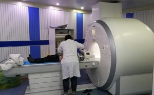
The new test is called MR Multitasking and tackles a common problem cardiologists face which is getting a still image when the heart is pumping and blood is flowing, causing blurry images.
“It is challenging to obtain good cardiac magnetic resonance images because the heart is beating incessantly and the patient is breathing, so the motion makes the test vulnerable to errors,” Shlomo Melmed, executive VP of academic affairs and the dean of the Cedars-Sinai medical faculty, said. “By novel approaches this this long-standing problem, this research team has found a unique solution to improve cardiac care for patients around the world for years to come.”
Patients who have MRIs currently have to hold their breath for a brief moment while an image is taken. MRI technicians have to time the images to a specific part of the heartbeat, according to the researchers. One of the biggest challenges with that method is that it has been shown to be unreliable and unsuitable for patients who have irregular heartbeats or breathing problems.
The researchers instead incorporated the natural movements of the body in order to make the MR Multitasking unit.
“Our solution is like making a video instead of a still image,” Anthony G. Christodoulou, a research scientist and the study’s first author, said. “MR Multitasking continuously acquires image data and then, when the test is completed, the program separates out the overlapping sources of motion and other changes into multiple time dimensions.”
In a test, 10 healthy volunteers and 10 cardiac patients had an MRI using MR Multitasking. The results showed that it was accurate and increased comfort for patients because they no longer had to hold their breath. The researchers performed three cardiac MRI tests in 90 seconds, a much shorter time than standard procedures.
“Now we collect all the data throughout the entire test and sort it afterwards,” Debiao Li, one of the researchers on the study, said. “We get full control after the test, as opposed to trying to control the body’s natural movement during imaging.”
The researchers also reported that MR Multitasking creates images that are six-dimensional because it uses time and motion.
“If a picture is @D, then a video is 3D because it adds the passage of time,” Christodoulou said. “Our videos are 6D because we can play them back four different ways. We can playback cardiac motions, respiratory motion and two different tissue processes that reveal cardiac health.”
MR Multitasking is going through clinical investigation at a number of medical centers throughout the U.S. and the researchers hope to expand the process to other diseases like cancer.
“People have to breath no matter what disease they have, so the ability to separate out the motion is important to many medical specialties,” Li said.
The research was published in the journal Nature Biomedical Engineering and was support by grants from the National Institutes of Health.