Medical Design and Outsourcing

  • Home
  • Medical Device Business
    • Mergers & Acquisitions
    • Financial
    • Regulatory
  • Applications
    • Cardiovascular
    • Devices
    • Imaging
    • Implantables
    • Medical Equipment
    • Orthopedic
    • Surgical
  • Technologies
    • Supplies and Components Index
    • Contract Manufacturing
    • Components
    • Electronics
    • Extrusions
    • Materials
    • Motion Control
    • Prototyping
    • Pumps
    • Tubing
  • MedTech Resources
    • Medtech Events in 2025
    • The 2024 Medtech Big 100
    • Medical Device Handbook
    • MedTech 100 Index
    • Subscribe to Print Magazine
    • DeviceTalks
    • Digital Editions
    • eBooks
    • Educational Assets
    • Manufacturer Search
    • Podcasts
    • Print Subscription
    • Webinars / Digital Events
    • Whitepapers
    • Voices
    • Video
  • 2025 Leadership
    • 2024 Winners
    • 2023 Winners
    • 2022 Winners
    • 2021 Winners
  • Women in Medtech
  • Advertise
  • Subscribe

Using data science to achieve ultra-low dose CT image reconstruction

August 25, 2015 By MDO Editor

Prof. Jeff Fessler and Prof. Yong Long, Ph.D. of the University of Michigan-Shanghai Jiao Tong University (UM-SJTU) Joint Institute are collaborating on a project to develop a dramatically improved approach to low-dose X-ray CT image formation by extracting and using information from a big-data corpus of regular dose X-Ray Computed Tomography (CT) images. The research is funded through the UM-SJTU Collaborative Research Programs for Energy and Biomedical Technology, which funds projects that have future commercial potential.

CT provides high-resolution images of anatomical structures for diagnosis and management of human diseases and disorders. It has already had a tremendous impact on cancer. For example, CT data are used routinely to help detect abnormal growths, diagnose tumors, plan treatment for radiation oncology, guide biopsy procedures, assess of tumor responses to treatment and monitor for recurrence.

Because of their value in patient care, there has been a dramatic increase in the number of CT scans ordered over the past several decades. In fact, CT scans are now are responsible for nearly 50% of the radiation exposure from medical imaging in the U.S. Unfortunately, the ionizing radiation in the form of X-rays used in CT scans is energetic enough to directly or indirectly damage DNA, putting patients at a higher risk for radiation-induced cancer. Studies have suggested that current CT usage may be responsible for 1.5-2% of all cancers in the U.S.

ultra-low-dose-img

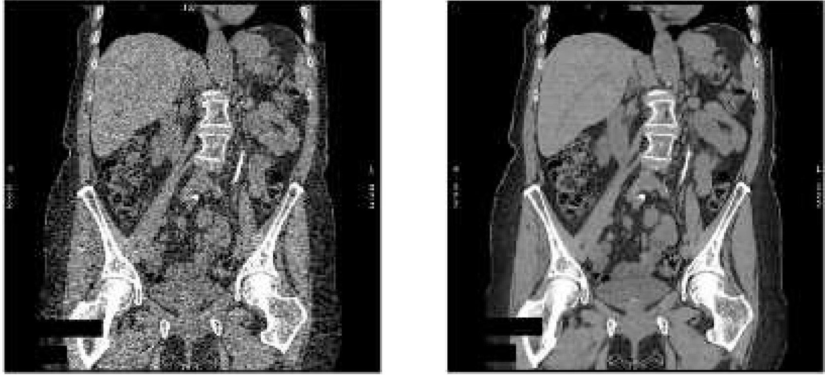
Coronal reformatted slice of 3D helical X-ray CT scan of the abdomen/pelvis. Reconstructed by the conventional FBP method (left) and by a MBIR method (right). For these thin-slice images, the MBIR method exhibits much lower noise than the FBP images. (Patient identifiers were removed.)

Ultra-low dose CT (ULDCT) scans that provide superior image quality could not only benefit patients who currently rely on the technology, but they could open up entirely new clinical applications. For example, the U.S. Preventive Services Task Force recommends annual screening for lung cancer with LDCT in high-risk adults. The same screening could be available to a wider population of adults if ultra-LDCT scans were available. This would help with detection and treatment of early-stage lung cancer and prevent a substantial number of lung cancer-related deaths, especially in China where lung cancer is the leading cause of cancer-related death. The death rate of lung cancer patients in China has increased by 465% over the past thirty years, and the number of individuals suffering from lung cancer in China could top one million by 2025.

Currently most commercial CT scanners use a technique called filter-back projection (FBP) for image reconstruction. However, FBP requires higher than desired doses of radiation to produce high-quality diagnostic images. Model-based image reconstruction (MBIR) methods, also known as statistical image reconstruction methods, improve the ability to produce high-quality and accurate images, while greatly reducing patient exposure to potentially harmful levels of radiation.

General Electrical introduced the world’s first commercial MBIR product, Veo, which allows CT scans to be performed using a radiation dose that is up to 75% lower than conventional image construction technique. It is currently in use at University of Michigan Hospital.

To achieve an even greater reduction in the radiation to achieve ULDCT scans, Fessler and Long plan to take an entirely new approach, one that makes use of the vast archive of existing CT images of patients with similar ailments.

University of Michigan-Shanghai Jiao Tong University
www.en.sjtu.edu.cn

University of Michigan
www.umich.edu

Related Articles Read More >

This is a Zimmer Biomet marketing image of its Rosa robotic surgery system.
Zimmer Biomet seeks a ZBEdge for its Rosa robotic surgery
A photo of Capstan Medical's mitral valve implant, which uses nitinol.
Capstan Medical’s R&D head discusses the heart valve and robotics startup’s tech, engineering challenges and solutions, advice for others in medtech and how to join his team
An illustration of a neurosurgeon using a robotic endoscope to remove a brain tumor.
MDO Nitinol Innovation Special Report
A photo of Highridge Medical CEO Rebecca Whitney.
Highridge Medical is betting on this spine tech
“mdo
EXPAND YOUR KNOWLEDGE AND STAY CONNECTED
Get the latest medical device business news, application and technology trends.

DeviceTalks Weekly

See More >

MDO Digital Edition

Digital Edition

Subscribe to Medical Design & Outsourcing. Bookmark, share and interact with the leading medical design engineering magazine today.

MEDTECH 100 INDEX

Medtech 100 logo
Market Summary > Current Price
The MedTech 100 is a financial index calculated using the BIG100 companies covered in Medical Design and Outsourcing.
DeviceTalks

DeviceTalks is a conversation among medical technology leaders. It's events, podcasts, webinars and one-on-one exchanges of ideas & insights.

DeviceTalks

New MedTech Resource

Medical Tubing

MassDevice

Mass Device

The Medical Device Business Journal. MassDevice is the leading medical device news business journal telling the stories of the devices that save lives.

Visit Website
MDO ad
Medical Design and Outsourcing
  • MassDevice
  • DeviceTalks
  • MedTech100 Index
  • Medical Tubing + Extrusion
  • Medical Design Sourcing
  • Drug Delivery Business News
  • Drug Discovery & Development
  • Pharmaceutical Processing World
  • R&D World
  • About Us/Contact
  • Advertise With Us
  • Subscribe to Print Magazine
  • Subscribe to our E-Newsletter
  • Listen to our Weekly Podcasts
  • Join our DeviceTalks Tuesdays Discussion

Copyright © 2025 WTWH Media, LLC. All Rights Reserved. The material on this site may not be reproduced, distributed, transmitted, cached or otherwise used, except with the prior written permission of WTWH Media LLC. Site Map | Privacy Policy | RSS

Search Medical Design & Outsourcing

  • Home
  • Medical Device Business
    • Mergers & Acquisitions
    • Financial
    • Regulatory
  • Applications
    • Cardiovascular
    • Devices
    • Imaging
    • Implantables
    • Medical Equipment
    • Orthopedic
    • Surgical
  • Technologies
    • Supplies and Components Index
    • Contract Manufacturing
    • Components
    • Electronics
    • Extrusions
    • Materials
    • Motion Control
    • Prototyping
    • Pumps
    • Tubing
  • MedTech Resources
    • Medtech Events in 2025
    • The 2024 Medtech Big 100
    • Medical Device Handbook
    • MedTech 100 Index
    • Subscribe to Print Magazine
    • DeviceTalks
    • Digital Editions
    • eBooks
    • Educational Assets
    • Manufacturer Search
    • Podcasts
    • Print Subscription
    • Webinars / Digital Events
    • Whitepapers
    • Voices
    • Video
  • 2025 Leadership
    • 2024 Winners
    • 2023 Winners
    • 2022 Winners
    • 2021 Winners
  • Women in Medtech
  • Advertise
  • Subscribe