 Researchers from the University of Texas Southwestern Medical Center have developed a threshold nanosensor that can differentiate between cancerous cells and healthy tissue during surgery. The team’s work was published in Nature Biomedical Engineering.
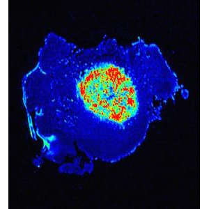
“We synthesized an imaging probe that stays dark in normal tissues but switches on like a light bulb when it reaches solid tumors. The purpose is to allow surgeons to see tumors better during surgery,” co-senior author Jinming Gao said in prepared remarks.
The nanosensor measures pH, or acid level, in a patient’s tissue and reacts according to the measured concentration. “Cancer is a very diverse set of diseases, but it does have some universal features,” co-senior author Dr. Baran Sumer explained. “Tumors do not have the same pH as normal tissue. Tumors are acidic, and they secrete acids into the surrounding tissue. It’s a very consistent difference and was discovered in the 1920’s.”
The researchers hope the new technology can reduce false rates in imaging and assist in non-invasive monitoring of drug responses.
“This new digital nanosensor-guided surgery potentially has several advantages for patients, including more accurate removal of tumors, and greater preservation of functional normal tissues,” Sumer said. “These advantages can improve both survival and quality of life.”