[Image from Stanford University]
“I’m using 3D-printed tools to design cardiac-mapping catheters, device used by surgeons to map the electrical activity of the heart and find disturbances,” Kevin Cyr, a second-year medical student at the university, said in a press release.
Atrial fibrillation (AFib) is a heart condition characterized by an irregular heartbeat that can eventually lead to blood clots, stroke, heart failure and other heart-related complications. According to the American Heart Association, about 2.7 million Americans are currently living with the condition.
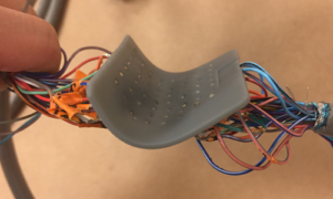
Cyr suggests the finding and understanding the rhythm disturbances that happen in the heart could be challenging because most of the medical devices that are typically used, which use electrodes on the surface of the heart to measure electrical activity, are one-size-fits-all. So, Stanford researchers are creating devices that can be customized to fit individual patients’s unique contours and divots of the heart.
In order for Cyr’s device to work, patients have to undergo an MRI or CT scan that records an image file of the heart which is then sent to a 3D printer. Using the scan, the researchers can recreate the natural anatomy of each patient and apply that knowledge to the device.
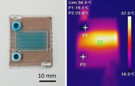
The device is small, thin and features a flexible silicone membrane with tiny holes in a grid-like formation. Each hole contains a tiny electrode that when placed on the surface of the heart’s atrium, can measure the electrical activity on that specific region of the heart. The data collected from the electrode can be transmitted to a computer creates a recording that shows the electrical activity of that area of the heart. The recordings also show a heat map of electrical activity that physicians can use to determine which areas of the heart need treatment.
“We can map in perfect detail this rectangular grid of information and not have to worry about missing signals, poor contact or things like that, which otherwise might throw out errors,” Cyr said.
Currently, the devices are being used on the epicardium layer of the heart. The researchers are working on seeing if the 3D-printed device could be used to map the interior of the heart as well. Using recordings from the interior of the heart could bring more accuracy in measuring rhythmic disturbance, according to the researchers.
The researchers suggest that it could take another year or two to refine the technology. It has not yet been tested on humans.