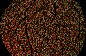
Using fluorescent protein markers and high-resolution imaging, researchers can visualize individual nerve fibers, show how different fiber types are organized within the nerve bundles and learn what kinds of information each group of fibers carries. [Image courtesy of Feinstein Institutes]
“This dataset represents a major step forward in bioelectronic medicine, offering the most detailed anatomical reconstruction of the human vagus nerve to date,” project leader Dr. Stavros Zanos said in a news release. “For the first time, we can visualize the vagus nerve’s complex architecture that will allow us to design more precise, effective and safe neuromodulation therapies and devices.”
The vagus nerve is the longest cranial nerve. It consists of two main bundles containing over 200,000 individual nerve fibers, stretching from the brainstem to all major organs, controlling autonomic nervous system functions like heart rate, breathing, digestion, immune response and inflammation.
“Even though much is known about the macroscopic and microscopic anatomy of the vagus, the spatial organization of fascicles and fibers within the nerve, as it relates to the innervated organs and the sensory and motor functions of the vagus, is largely unknown,” the study’s purpose said.
Funded by a $6.7 million National Institutes of Health (NIH) grant, the researchers sought to create a comprehensive anatomical map to share with the scientific community to increase understanding of the autonomic nervous system and advance vagus neuromodulation therapies.
Over three years, researchers created the anatomical map by analyzing 60 vagus nerves from 30 human donors. They used microCT imaging, immunohistochemistry and ultrasound to offer a 3D view into the vagus nerve and better understand the function of each vagal fiber.
After completing the first data set, the researchers published their findings online to help others in the scientific community. Others could view ultrasound images and video of the vagus nerve, microCT scans across the length of the nerve and images across multiple levels of the nerve.
Related: Researchers test microrobots for treating spinal cord injuries
With the comprehensive map, researchers could gain insight into how the vagus nerve communicates with organs and impacts health and disease through the organization of fascicles and fibers. The study is ongoing, as the recent data published consists of one dataset. Additional data is expected to be uploaded in the future and the researchers said they’ll publish initial conclusions with more sufficient data.
“Decoding the vagus nerve’s intricate language is an important advance for science and medicine,” Feinstein Institutes President and CEO Dr. Kevin Tracey said. “This knowledge will further empower researchers to re-engineer human biology and unlock novel therapies for future patients.”
Tracey discovered how the brain and body communicate through the vagus nerve to regulate inflammation over 30 years ago. His findings led to the development of the first FDA-approved vagus nerve stimulation device to treat rheumatoid arthritis, the SetPoint system. The device was first implanted in patients in August 2025.